
🩻 🩻
Bone Densitometer (DEXA Scanner): How It Works, Applications, and Future of Osteoporosis Diagnosis
Table of Contents
- What Is a Bone Densitometer (DEXA Scanner)
- A Short History of Bone Densitometry
- How It Works: The Imaging Principle
- Key Hardware Components
- Clinical Applications & Use Cases
- DEXA vs Other Imaging Modalities
- Safety & Radiation Dose
- Maintenance & Technical Considerations (for Biomedical Engineers)
- Cost & Accessibility
- Future Innovations & Trends
- Simple Analogies to Demystify DEXA
- Final Summary
- References
1. What Is a Bone Densitometer (DEXA Scanner)
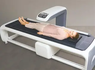
A Bone Densitometer, also known as a DEXA Scanner (Dual-Energy X-ray Absorptiometry), is a specialized medical imaging device designed to measure Bone Mineral Density (BMD).
It is the gold standard for diagnosing osteoporosis, a condition where bones become weak and fragile, increasing fracture risk.
Unlike conventional X-rays that visualize bone structure, a DEXA scanner quantifies bone strength, allowing clinicians to detect bone loss even before a fracture occurs.
2. A Short History of Bone Densitometry
The evolution of bone densitometry reflects advances in precision imaging:
- 1960s: Introduction of Single-Photon Absorptiometry (SPA) — the earliest bone density measurement technique.
- 1970s–1980s: Development of Dual-Energy X-ray Absorptiometry (DEXA), which improved accuracy and reduced radiation.
- 1990s: Commercialization by leading manufacturers such as GE Lunar and Hologic, establishing DEXA as the clinical standard.
- 2000s–Present: Digital imaging, automated reporting, and AI-based fracture risk prediction have modernized bone health assessment.
3. How It Works: The Imaging Principle
DEXA uses two low-dose X-ray beams with different energy levels.
As these beams pass through bone and soft tissue, they are absorbed differently.
By measuring this differential absorption, the system calculates the bone mineral content (BMC) and bone mineral density (BMD).
🧠 In simple terms:
It’s like shining two colored flashlights (for example, red and blue) through an object.
If one color is absorbed more than the other, you can estimate how dense that object is — this is the same logic DEXA uses to measure bone density.
4. Key Hardware Components
| Component | Function |
|---|---|
| X-ray Source | Generates dual-energy beams (typically 70–140 kVp). |
| Detector Array | Captures transmitted beams to measure energy attenuation. |
| Patient Table | Smoothly moves the patient through the scanning path. |
| Calibration Phantom | Used to ensure accuracy and consistency of results. |
| Computer & Software | Processes data, calculates T-scores/Z-scores, and produces diagnostic reports. |
5. Clinical Applications & Use Cases
- Diagnosis of Osteoporosis and Osteopenia
- Monitoring Response to Treatment (e.g., bisphosphonates, vitamin D therapy)
- Fracture Risk Prediction
- Body Composition Analysis (fat, lean mass, and muscle distribution)
- Pediatric Bone Studies (growth and development monitoring)
6. DEXA vs Other Imaging Modalities
| Imaging Modality | Primary Use | Advantages | Limitations |
|---|---|---|---|
| DEXA | Bone mineral density measurement | Accurate, fast, very low dose | Limited 3D detail |
| QCT (Quantitative CT) | Volumetric bone analysis | 3D imaging, precise geometry | Higher radiation, costly |
| Ultrasound (QUS) | Bone quality estimation | Portable, no radiation | Lower precision, site dependent |
| Plain X-ray | Detects fractures | Widely available | Cannot measure bone density |
DEXA offers the best balance of accuracy, safety, and cost, which explains why it’s the clinical gold standard.
7. Safety & Radiation Dose
DEXA scanners are among the lowest-radiation imaging systems in medicine.
A standard DEXA scan exposes the patient to 0.001–0.005 mSv, roughly equivalent to one day of natural background radiation.
Modern DEXA units employ:
- Beam collimation to focus exposure only on the region of interest.
- Automatic exposure control (AEC) for dose optimization.
- Lead shielding and interlock systems for operator safety.
8. Maintenance & Technical Considerations (for Biomedical Engineers)
Biomedical engineers play a vital role in ensuring accurate results and system reliability.
Key maintenance tasks include:
- Daily or weekly phantom calibration to maintain accuracy.
- Regular tube output and detector uniformity checks.
- Mechanical alignment verification of the scanning arm.
- Ensuring software updates comply with WHO and ISCD standards.
Troubleshooting tip:
If bone density results fluctuate significantly (>1–2%) across repeated scans, calibration or detector drift may be the cause.
9. Cost & Accessibility
| System Type | Typical Price (USD) | Ideal Setting |
|---|---|---|
| Portable DEXA (forearm/heel) | $20,000–$40,000 | Clinics, mobile screening |
| Standard Tabletop System | $45,000–$70,000 | General hospitals |
| Advanced Whole-Body DEXA | $80,000–$130,000 | Research centers, academic hospitals |
Operating costs are minimal — power consumption and periodic calibration are the main expenses.
10. Future Innovations & Trends
- AI-Based Bone Fracture Prediction: Machine learning algorithms analyzing trabecular patterns.
- 3D Bone Density Mapping: Integration with CT and MRI for comprehensive skeletal models.
- Portable Handheld DEXA: Emerging devices for bedside and community screening.
- Cloud-Based PACS Integration: For tele-radiology and data sharing.
- Automated Body Composition Analytics: Used in sports medicine and obesity research.
The future of DEXA lies in combining precision imaging with predictive analytics — transforming how bone health is managed globally.
11. Simple Analogies to Demystify DEXA
Think of your bones like reinforced concrete — calcium and minerals are the “cement.”
A DEXA scanner doesn’t just photograph the concrete; it measures how much cement is left inside.
This tells doctors whether the bone is strong enough to withstand daily stress.
12. Final Summary
- The Bone Densitometer (DEXA) is the gold standard for osteoporosis diagnosis.
- It works by measuring the absorption difference of two X-ray beams through bone and soft tissue.
- DEXA is safe, accurate, and cost-effective, with very low radiation exposure.
- Biomedical engineers must maintain calibration, alignment, and QA records to ensure precision.
- Future systems will rely heavily on AI, cloud computing, and 3D analytics.
13. References
Njeh CF et al. Comparative Analysis of Bone Densitometry Techniques. Phys Med Biol, 2019.
World Health Organization (WHO). Prevention and Management of Osteoporosis: Technical Report Series 921.
International Society for Clinical Densitometry (ISCD), Official Positions 2023.
Blake GM, Fogelman I. The Role of DXA in Osteoporosis Diagnosis. Bone, 2007.
Hologic & GE Healthcare DEXA System Datasheets.