Understanding the Anatomy and Physiology of the Heart: A Complete Biomedical Guide for Students and Clinicians
Introduction
The human heart is one of the most important organs in the body. It works continuously from before birth until death, pumping blood to every tissue and organ. A healthy heart delivers oxygen, nutrients, hormones, and immune cells throughout the body while removing waste products such as carbon dioxide.
In simple terms, the heart acts like a double pump:
- the right side pumps blood to the lungs
- the left side pumps blood to the body
Understanding the anatomy and physiology of the heart is essential for:
- medical students
- biomedical engineering students
- nursing learners
- anatomy and physiology beginners
This article explains the structure and function of the heart in a simplified academic style, connecting anatomy with physiology for easier learning and memory retention.
Table of Contents
- Introduction
- Overview of the Cardiovascular System
- Location and External Anatomy of the Heart
- Layers of the Heart Wall
- Chambers of the Heart
- Heart Valves
- Blood Flow Through the Heart
- Coronary Circulation
- Electrical Conduction System
- Cardiac Cycle
- Physiology of Cardiac Muscle
- Cardiac Output and Blood Pressure
- Regulation of Heart Function
- Clinical Relevance
- Conclusion
- Key Takeaways
- Academic References
1. Overview of the Cardiovascular System
The cardiovascular system consists of:
- the heart
- blood vessels
- blood
Its major functions include:
- transporting oxygen and nutrients
- removing metabolic waste
- maintaining body temperature
- supporting immune defense
- distributing hormones
The heart is the central organ of this system.
A useful analogy is to think of the cardiovascular system as a city’s water supply network:
- the heart = pumping station
- arteries = delivery pipes
- veins = return pipes
- blood = transport fluid
2. Location and External Anatomy of the Heart
The heart is located in the thoracic cavity between the lungs in a region called the mediastinum. It lies slightly to the left of the midline behind the sternum.
Basic Characteristics
| Feature | Description |
|---|---|
| Size | Approximately the size of a fist |
| Weight | 250–350 grams |
| Shape | Cone-shaped muscular organ |
| Position | Between the lungs |
| Apex | Points downward and left |
| Base | Faces upward and backward |
Major External Structures
- Aorta
- Pulmonary trunk
- Superior vena cava
- Inferior vena cava
- Pulmonary veins
- Coronary arteries
Simple Memory Tip
“Apex points Left.”
This helps students remember the direction of the heart’s lower tip.
3. Layers of the Heart Wall
The heart wall contains three major layers.
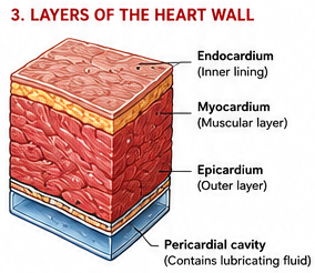
1. Endocardium
- Inner lining
- Smooth epithelial layer
- Reduces friction during blood flow
2. Myocardium
- Thick muscular middle layer
- Responsible for contraction
- Thickest in the left ventricle
3. Epicardium
- Outer protective layer
- Part of the pericardium
4. Pericardium
The heart is enclosed within a protective sac called the pericardium.
Functions of the Pericardium
- protects the heart
- reduces friction
- anchors the heart
- prevents overexpansion
Pericardial Layers
| Layer | Function |
|---|---|
| Fibrous pericardium | Tough outer protection |
| Serous pericardium | Produces lubricating fluid |
Between the layers is the pericardial cavity, containing fluid that reduces friction during heartbeats.
5. Chambers of the Heart
The heart has four chambers.
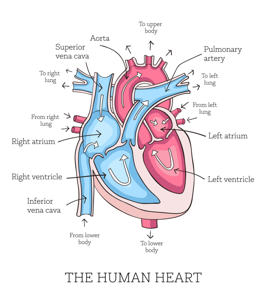
| Chamber | Function |
|---|---|
| Right atrium | Receives deoxygenated blood |
| Right ventricle | Pumps blood to lungs |
| Left atrium | Receives oxygenated blood |
| Left ventricle | Pumps blood to body |
Important Concept
The:
- atria receive blood
- ventricles pump blood out
The left ventricle has the thickest wall because it must pump blood throughout the entire body.
Easy Analogy
Think of:
- atria = waiting rooms
- ventricles = powerful pumping chambers
6. Heart Valves
Heart valves ensure one-way blood flow.
The Four Heart Valves
| Valve | Location | Function |
|---|---|---|
| Tricuspid valve | RA → RV | Prevents backflow |
| Pulmonary valve | RV → pulmonary artery | Controls flow to lungs |
| Mitral valve | LA → LV | Prevents backflow |
| Aortic valve | LV → aorta | Controls flow to body |
Memory Trick
“Try Pulling My Aorta”
- Tricuspid
- Pulmonary
- Mitral
- Aortic
Valves open and close because of pressure changes.
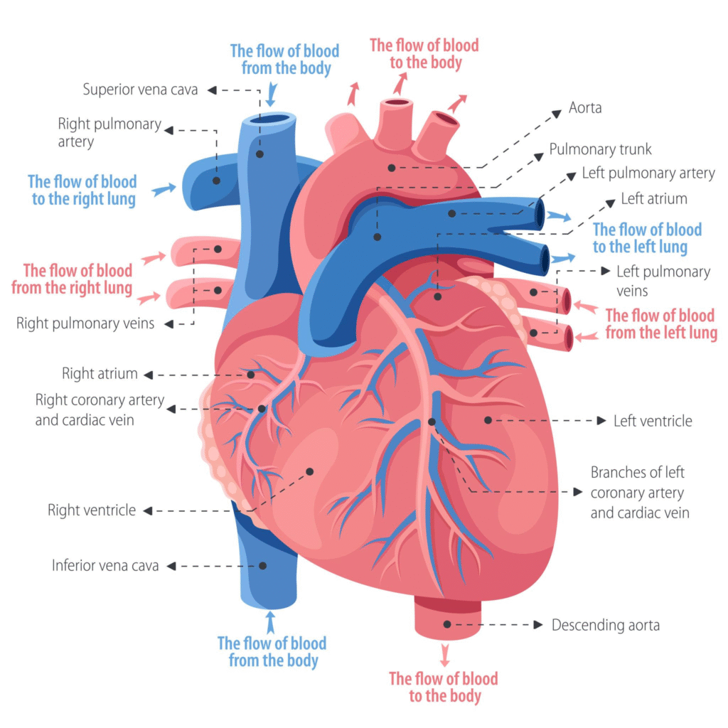
7. Blood Flow Through the Heart
Understanding blood flow is one of the most important concepts in cardiac anatomy.
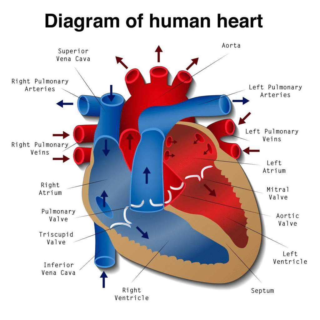
Blood Flow Pathway
- Body tissues
- Superior and inferior vena cava
- Right atrium
- Tricuspid valve
- Right ventricle
- Pulmonary valve
- Pulmonary arteries
- Lungs
- Pulmonary veins
- Left atrium
- Mitral valve
- Left ventricle
- Aortic valve
- Aorta
- Body tissues
Pulmonary vs Systemic Circulation
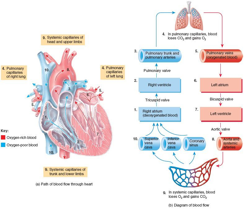
| Type | Pathway | Purpose |
|---|---|---|
| Pulmonary circulation | Heart ↔ lungs | Gas exchange |
| Systemic circulation | Heart ↔ body | Deliver oxygen |
8. Coronary Circulation
The heart muscle itself requires oxygen and nutrients. These are supplied by the coronary arteries.
Main Coronary Arteries
- Right coronary artery (RCA)
- Left coronary artery (LCA)
The LCA divides into:
- Left anterior descending artery (LAD)
- Circumflex artery
Clinical Importance
Blocked coronary arteries can cause:
- angina
- myocardial infarction (heart attack)
A heart attack occurs when blood flow to heart muscle becomes blocked.
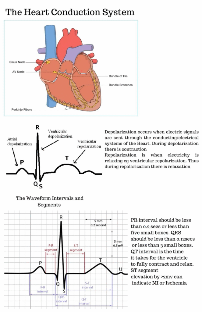
9. Electrical Conduction System
The heart can generate its own electrical impulses. This system coordinates heartbeat rhythm.
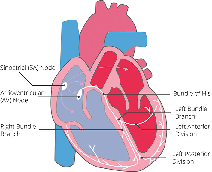
Main Components
| Structure | Function |
|---|---|
| SA node | Natural pacemaker |
| AV node | Delays impulse |
| Bundle of His | Conducts impulses |
| Bundle branches | Carry signals |
| Purkinje fibers | Stimulate ventricles |
Sequence of Electrical Activity
- SA node fires
- Atria contract
- AV node delays signal
- Ventricles contract
ECG Connection
Electrical activity can be recorded using an electrocardiogram (ECG).
| ECG Wave | Meaning |
|---|---|
| P wave | Atrial depolarization |
| QRS complex | Ventricular depolarization |
| T wave | Ventricular repolarization |
10. Cardiac Cycle
The cardiac cycle refers to one complete heartbeat.
It includes:
- contraction (systole)
- relaxation (diastole)
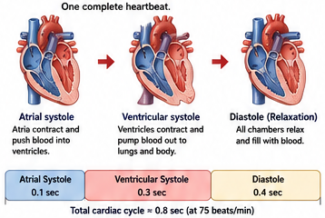
Important Definitions
| Term | Meaning |
|---|---|
| Heart rate (HR) | Beats per minute |
| Stroke volume (SV) | Blood pumped per beat |
| Cardiac output (CO) | Blood pumped per minute |
Normal cardiac output is approximately 5 L/min in adults.
11. Physiology of Cardiac Muscle
Cardiac muscle is unique.
Characteristics of Cardiac Muscle
- involuntary
- striated
- branched
- highly aerobic
- rich in mitochondria
Intercalated Discs
Cardiac cells connect through structures called intercalated discs.
These contain:
- gap junctions
- desmosomes
They allow rapid electrical communication.
Why the Heart Does Not Tire Easily
The heart contains many mitochondria and receives constant oxygen supply.
This allows continuous ATP production.
12. Action Potentials in the Heart
Cardiac action potentials differ from nerve action potentials.
Phases of Cardiac Action Potential
| Phase | Event |
|---|---|
| 0 | Rapid depolarization |
| 1 | Initial repolarization |
| 2 | Plateau phase |
| 3 | Repolarization |
| 4 | Resting potential |
The plateau phase is important because it prevents tetanic contractions.
This ensures the heart can relax between beats.
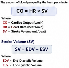
13. Cardiac Output and Blood Pressure
Cardiac output measures how effectively the heart pumps blood.
SV = EDV – ESV
Where:
- EDV = end-diastolic volume
- ESV = end-systolic volume
Factors Affecting Cardiac Output
1. Heart Rate
Faster heartbeat can increase output.
2. Stroke Volume
Greater ventricular filling increases pumping force.
3. Contractility
Stronger contraction increases output.
14. Regulation of Heart Function
The autonomic nervous system regulates the heart.
| Division | Effect |
|---|---|
| Sympathetic | Increases heart rate |
| Parasympathetic | Decreases heart rate |
Hormones such as adrenaline also affect heart activity.
15. Clinical Relevance
Understanding heart anatomy and physiology is essential in medicine.
Common Heart Diseases
| Disease | Description |
|---|---|
| Coronary artery disease | Blocked arteries |
| Heart failure | Weak pumping ability |
| Arrhythmia | Abnormal rhythm |
| Valve disease | Valve malfunction |
| Hypertension | High blood pressure |
Heart Failure
Heart failure occurs when the heart cannot pump effectively.
Symptoms
- fatigue
- shortness of breath
- edema
- exercise intolerance
Arrhythmias
Problems in the electrical conduction system may cause irregular heart rhythms.
Examples:
- atrial fibrillation
- ventricular tachycardia
- heart block
Conclusion
The heart is an extraordinary organ that combines complex anatomy with highly coordinated physiology. Its chambers, valves, muscle layers, blood vessels, and electrical system work together continuously to maintain life.
For students of anatomy and physiology, understanding the relationship between structure and function is the key to mastering cardiovascular science. The anatomy explains where structures are located, while physiology explains how they function together.
A strong understanding of heart anatomy and physiology also forms the foundation for learning:
- cardiology
- pathology
- biomedical engineering
- pharmacology
- clinical medicine
The heart is not simply a pump. It is a dynamic biological engine precisely regulated to meet the body’s changing demands every second of life.
Key Takeaways
- Heart has 4 chambers.
- Atria receive blood; ventricles pump blood.
- Left ventricle has the thickest myocardium.
- Four valves ensure one-way blood flow.
- SA node is the natural pacemaker.
- Coronary arteries supply the heart muscle.
- Cardiac cycle includes systole and diastole.
- Cardiac output = heart rate × stroke volume.
- Pulmonary circulation goes to lungs.
- Systemic circulation goes to body.
- Heart diseases often involve arteries, valves, or electrical conduction.
Academic References
- Oberman, R., Shumway, K. R., & Bhardwaj, A. (2023). Physiology, Cardiac. StatPearls Publishing. NCBI Bookshelf.
NCBI Bookshelf – Physiology, Cardiac
(ncbi.nlm.nih.gov) - Ripa, R., George, T., Shumway, K. R., & Sattar, Y. (2023). Physiology, Cardiac Muscle. StatPearls Publishing.
NCBI Bookshelf – Cardiac Muscle Physiology
(ncbi.nlm.nih.gov) - Rehman, I., & Rehman, A. (2023). Anatomy, Thorax, Heart. StatPearls Publishing. PubMed.
PubMed – Anatomy, Thorax, Heart
(PubMed) - Mayo Clinic – Heart Disease Overview
(Mayo Clinic) - Mayo Clinic – Heart Attack Overview
(Mayo Clinic) - Cleveland Clinic – Heart Anatomy & Function
(Cleveland Clinic) - Hall, J. E. (2020). Guyton and Hall Textbook of Medical Physiology (14th ed.). Elsevier.
Elsevier – Guyton and Hall Textbook - Standring, S. (2020). Gray’s Anatomy: The Anatomical Basis of Clinical Practice (42nd ed.). Elsevier.
Elsevier – Gray’s Anatomy - NIH National Heart, Lung, and Blood Institute
- World Health Organization – Cardiovascular Diseases