
How Do Surgical Lasers Work? Wavelengths, Tissue Interaction, and Biomedical Engineering Guide
A surgical laser is a device that focuses a beam of a single wavelength of light onto tissue to cut, vaporize, or coagulate it — with no mechanical blade involved. What a given laser does to tissue is set almost entirely by its wavelength: a wavelength that water absorbs strongly (like the 10,600 nm CO₂ laser) stays superficial and vaporizes tissue cleanly, while a wavelength water absorbs weakly (like the 1,064 nm Nd:YAG laser) penetrates several millimeters deep and coagulates rather than cuts. In engineering terms, a surgical laser system is a precisely controlled energy-delivery instrument: an active medium that generates the beam, an optical or fiber path that delivers it to the tissue, and a control console that sets power, pulse duration, and repetition rate to hit a specific tissue effect.
Everything below unpacks that definition layer by layer — starting from why lasers replaced (and complement) the scalpel, through to how a system is built, operated, tested, maintained, and where the field is going next.
Table of contents
- 1. Why surgery needs a light-based cutting tool
- 2. Working principle: wavelength decides the tissue effect
- 3. Device architecture and components
- 4. Classification: laser hazard class, not a device “type”
- 5. Control parameters: what the surgeon actually sets
- 6. Step-by-step: a typical laser procedure
- 7. Delivery fiber and power source considerations
- 8. QA, testing, and acceptance criteria
- 9. Complications, safety, and failure modes — with real numbers
- 10. Maintenance and troubleshooting
- 11. Regulatory and standards framework
- 12. Manufacturer landscape
- 13. Wavelength comparison at a glance
- 14. Future directions
- 15. Conclusion
- FAQ
- References
1. Why surgery needs a light-based cutting tool
A scalpel cuts by mechanical shear — it separates tissue but does nothing to stop bleeding, and it cannot vaporize tissue selectively. Electrosurgery (radiofrequency current) solves the bleeding problem but still requires tissue contact and can char a wide margin. A laser solves both limitations at once because it deposits energy optically, without touching the tissue:
- No-touch cutting — the beam can be aimed through air, through an endoscope’s working channel, or down an optical fiber threaded inside a ureteroscope, reaching sites a blade cannot.
- Wavelength-selective tissue effect — because different tissue chromophores (water, hemoglobin, melanin) absorb different wavelengths at very different rates, choosing the wavelength chooses the depth and type of damage — vaporization, cutting, or coagulation — before the surgeon even fires the laser.
- Simultaneous hemostasis — most surgical wavelengths coagulate small vessels as they cut, which is why lasers dominate in vascular, richly perfused, or hard-to-access tissue (skin, prostate, airway, GI mucosa, kidney stones).
The trade-off is that a laser is a much more complex, less forgiving instrument than a scalpel: it requires wavelength-specific eyewear for everyone in the room, a fire-safety protocol, and calibrated power/pulse settings — this is why the regulatory and QA sections below carry as much weight as the physics.
2. Working principle: wavelength decides the tissue effect
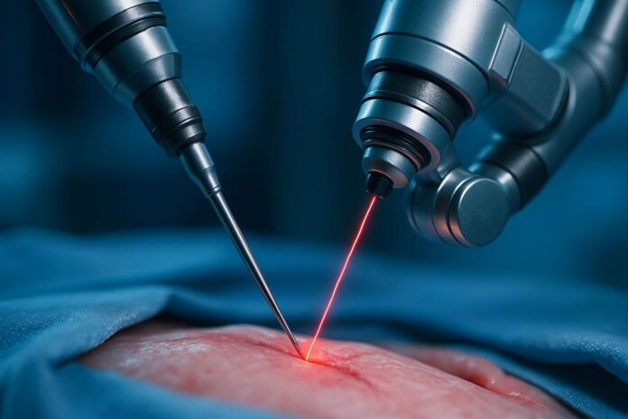
A laser (“Light Amplification by Stimulated Emission of Radiation”) produces coherent, single-wavelength light by pumping energy into an active medium (a gas, crystal, or semiconductor) until it emits photons in phase, in one direction, at one wavelength. What matters clinically is what happens next — how that wavelength interacts with tissue:
- Absorption depth — the number of micrometers or millimeters the light travels into tissue before roughly 90% of its energy has been absorbed. Water is the dominant absorber in soft tissue for most surgical wavelengths.
- Thermal spread — heat conducts outward from the absorption zone into adjacent tissue; shorter pulses and higher water absorption reduce this “collateral” zone, giving a more precise cut with less charring.
- Ablation vs. coagulation — enough energy delivered fast enough vaporizes tissue water explosively (ablation/cutting); slower or lower-energy delivery instead denatures proteins and seals vessels without vaporizing (coagulation).
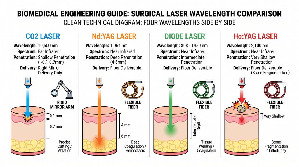
The four wavelengths used in most surgical lasers today, and what that wavelength buys clinically:
| Laser type | Wavelength | Water absorption | Typical tissue penetration | Dominant clinical effect |
|---|---|---|---|---|
| CO₂ | 10,600 nm (far-IR) | Very high | ~100–700 μm (superficial) | Precise vaporization/cutting, minimal thermal spread — skin, airway, general/plastic surgery |
| Nd:YAG | 1,064 nm (near-IR) | Low | 4–6 mm | Deep coagulation, can be fiber-delivered |
| Diode | 808–1,450 nm (near-IR) | Low–moderate | Intermediate, fiber-deliverable | Combined cutting + coagulation, compact solid-state systems |
| Ho:YAG | 2,100 nm (near-IR) | Very high | Very shallow, fiber-deliverable | Stone fragmentation (“vaporization/drilling”), soft-tissue cutting + coagulation in one device — endourology workhorse |
Note the pattern: CO₂ and Ho:YAG both sit at wavelengths water absorbs strongly, but CO₂ cannot travel down a flexible optical fiber (its far-infrared light is absorbed by standard silica fiber), so it is limited to line-of-sight or articulated-arm/waveguide delivery — while Ho:YAG’s shorter near-infrared wavelength travels down a flexible fiber, which is exactly why Ho:YAG, not CO₂, is the endoscopic stone-surgery laser.
3. Device architecture and components
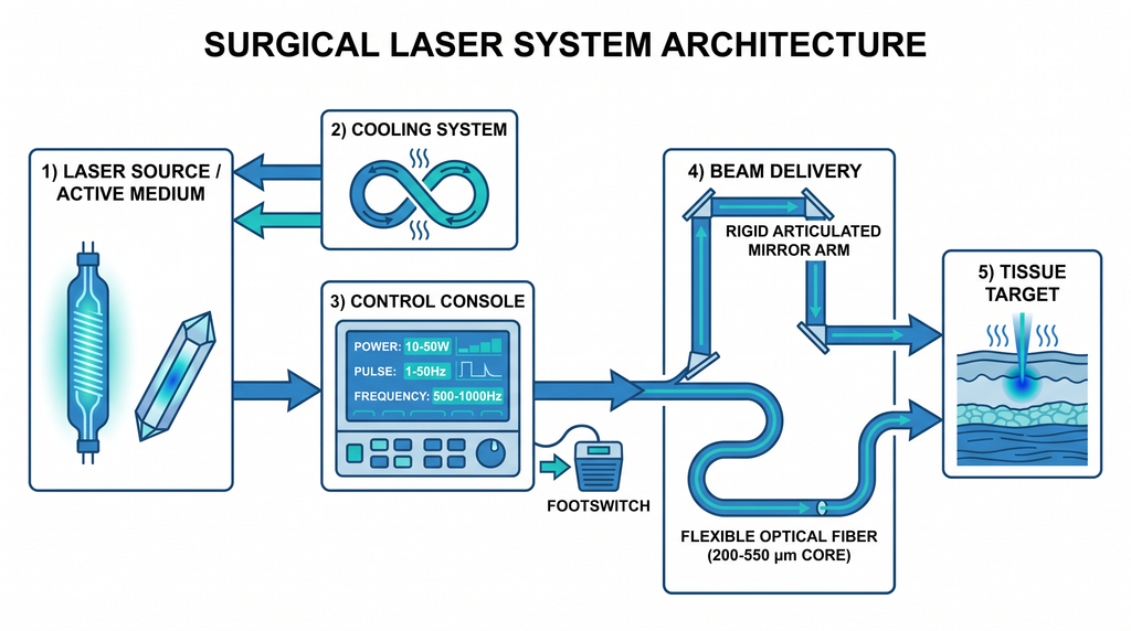
A surgical laser system has five functional blocks:
- Laser source / active medium — a sealed CO₂ gas tube (gas laser) or a solid-state crystal rod doped with a rare-earth ion (Nd:YAG = neodymium-doped yttrium aluminum garnet; Ho:YAG = holmium-doped version of the same crystal) pumped by a flashlamp or diode array.
- Beam delivery — either a rigid articulated arm with mirrors (required for CO₂, since its wavelength cannot travel through standard fiber) or a flexible optical fiber (200–550 μm core diameter for endoscopic Ho:YAG/diode/Nd:YAG systems).[^5] Smaller-core fibers (≤300 μm) are more maneuverable through narrow endoscope channels but show a measurably higher rate of connector-end failure than larger fibers — one MAUDE-database analysis found roughly a 4% connector failure rate for 200 μm fibers versus 0% for 365 μm fibers.
- Cooling system — closed-loop water or forced-air circulation around the active medium; without it the medium overheats and output power drifts or drops.
- Control console — sets average power (W), pulse energy (J), pulse duration, and repetition rate (Hz); shows real-time output on a display; and includes the safety interlocks described in Section 8.
- Footswitch / activation control — a dedicated foot pedal (never a handheld button) so the surgeon’s hands stay on the delivery device while firing, and firing only happens while the pedal is actively depressed.
4. Classification: laser hazard class, not a device “type”
Unlike some device families, surgical lasers don’t have a single clinical classification code — instead they carry a laser safety class that describes hazard, defined by IEC 60825-1 (Edition 3.0), harmonized with FDA guidance since Laser Notice No. 56 (February 2023):
- Class 1 — no hazard under normal use (fully enclosed systems only).
- Class 2 — visible-light, low-power (aiming beams only; not therapeutic).
- Class 3R — low-risk but capable of eye injury with direct viewing.
- Class 3B — hazardous to the eye from direct or specular reflection; diffuse reflections generally safe. Some lower-power therapeutic lasers fall here.
- Class 4 — hazardous to eye and skin from direct, reflected, or even diffusely scattered beams, and can ignite materials. Virtually all surgical CO₂, Nd:YAG, diode, and Ho:YAG systems are Class 4.
In the US, surgical lasers are separately regulated as medical devices under 21 CFR 878.4810 (“powered laser surgical instrument”), a Class II device requiring FDA 510(k) premarket clearance and reviewed under the General & Plastic Surgery panel.
5. Control parameters: what the surgeon actually sets
Every console exposes the same core parameters, and their combination — not any single number — determines the tissue effect:
- Average power (W) — total energy delivered per second; ranges from a few watts (fine dermatologic work) up to 60–120 W average, 240 W peak, on high-output CO₂ and Ho:YAG systems.
- Pulse energy (J) and pulse duration — how much energy each individual pulse carries and over what time; shorter, higher-peak-power pulses ablate more precisely with less thermal spread.
- Repetition rate (Hz) — pulses per second; commercial Ho:YAG lithotripsy systems commonly run 5–120 Hz depending on the model and stone-fragmentation mode.
- Spot size / fiber core diameter — sets power density (fluence) at the tissue surface for a given power setting; the same wattage through a smaller spot or fiber core produces a much higher local fluence.
Some modern Ho:YAG platforms add a pulse-shaping mode (marketed as e.g. MOSES technology) that splits each pulse into two phases — an initial low-energy pulse that displaces the surrounding fluid (“Moses effect”) before the main high-energy pulse arrives at the stone with less energy lost to the medium in between — improving fragmentation efficiency versus a plain single-pulse waveform.
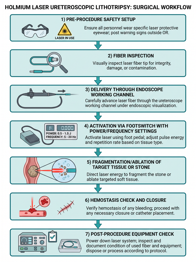
6. Step-by-step: a typical laser procedure
Using Ho:YAG ureteroscopic lithotripsy as a representative worked example (the same sequence generalizes to CO₂ skin resurfacing or diode soft-tissue work, with delivery method substituted):
- Pre-procedure safety setup — wavelength-specific protective eyewear on for every person in the room; laser warning sign posted on the door; for airway/CO₂ cases, flammable-prep and oxygen-concentration checks per fire-safety protocol.
- Fiber inspection — the optical fiber is visually inspected end-to-end for damage and confirmed to be firmly seated in the laser’s output aperture before insertion; a damaged or improperly seated fiber is the single most commonly cited equipment-related cause of laser adverse events in the literature (see Section 9).
- Delivery — the fiber (or, for CO₂, the articulated arm/handpiece) is passed through the endoscope’s working channel (or positioned over the target for open/topical use) under direct visualization; the fiber tip is confirmed in position and, for flexible-scope work, secured to prevent migration during the case.
- Activation — the surgeon sets power/frequency/pulse-energy parameters for the specific tissue task, then fires only via the dedicated footswitch, in short controlled bursts, watching the tissue response in real time and adjusting parameters between bursts.
- Fragmentation or ablation — for stone work, holmium energy vaporizes/drills through the stone into dust or small fragments, which are either allowed to pass spontaneously or retrieved with a basket; for soft-tissue work, the tissue surface vaporizes layer by layer under direct vision.
- Hemostasis check and closure — the treated field is inspected for residual bleeding (most surgical wavelengths self-coagulate small vessels; larger vessels may need supplemental treatment), any residual debris/fragments cleared, and the scope withdrawn or the field dressed.
- Post-procedure equipment check — fiber and console inspected for damage or performance drift before storage/reuse, per the manufacturer’s service protocol.
7. Delivery fiber and power source considerations
Fiber-delivered systems (Nd:YAG, diode, Ho:YAG) carry their own biomaterials-adjacent engineering constraints, distinct from CO₂’s rigid, mirror-based delivery:
- Core diameter trade-off — 200 μm fibers thread through narrower working channels and flex through tighter endoscope curves, but bend radii below about 0.6 cm significantly raise fracture risk versus larger-core fibers.
- Reusable vs. single-use fibers — reusable fibers require re-stripping and re-cleaving the tip between uses (tip degradation is a known source of reduced efficiency and increased proximal fiber heating); single-use fibers avoid this but add per-case cost.
- Power supply — these are line-powered systems (100–240 VAC), not battery devices; console footprints on commercial CO₂ platforms are on the order of 40 × 40 × 136 cm and draw up to roughly 9 A at mains voltage.
8. QA, testing, and acceptance criteria
Surgical laser systems require both incoming/periodic equipment QA and per-case checks:
- Power output verification — measured against the console’s displayed setting using a calibrated power meter; a mismatch beyond the manufacturer’s stated tolerance is an acceptance-test failure requiring service before further use.
- Beam alignment (for mirror/articulated-arm CO₂ systems) — the visible aiming beam must be confirmed coincident with the invisible treatment beam at the point of use; misalignment is a direct patient-safety hazard.
- Safety interlock function — door/room interlocks, footswitch dead-man behavior, and standby/ready-state indicators are tested on a defined schedule (commonly every 6–12 months, and per manufacturer service intervals) and before returning a serviced unit to clinical use.
- Fiber/connector inspection — pre-case visual inspection for cladding damage, tip degradation, or connector wear; smaller-core (≤300 μm) fibers warrant closer scrutiny given their higher documented connector-failure rate.
- Plume evacuation verification — adequate smoke evacuation airflow at the point of ablation, checked as part of routine OR equipment readiness, since laser plume carries particulate and potentially viable biological material.
9. Complications, safety, and failure modes — with real numbers
The best available real-world failure data comes from national/regional adverse-event registries and device-incident databases rather than single-center case series, because equipment failures are relatively rare per case but consequential in aggregate:
French national data (HAS database, urology, May 2016–December 2023, 149 laser-related care-associated adverse events out of 1,376 total CAEs reported):
- Severity: 92.6% were mild-to-moderate (Grade 1–2); 5 events (3.4%) were Grade 3 (e.g., significant bleeding requiring transfusion, sepsis requiring ICU care); 6 events (4%) were Grade 4 (life-threatening — including a cerebrovascular accident after ureteroscopy and urethro-rectal/urethro-pubic fistulae).
- Root cause distribution: material/equipment problems, 38.25% (the single largest cause); patient information issues, 24.83%; medications, 15.43%; technical/operator gesture, 14.1%; case clinical complexity, 7.38%.
- Material-related incidents were significantly more common in younger patients (p<0.001), healthier (ASA 1) patients (p=0.003), and urgent procedures (p<0.001) — the authors interpret this as material failure striking somewhat independently of patient risk factors, unlike most surgical complication categories.
US MAUDE database, laser fibers and generators in ureteroscopy/lithotripsy, 10-year analysis (2024):
- The most common equipment problems were fiber breakage outside the patient during use (26.3%) and breakage of the fiber tip (21.2%), followed by generator malfunction and connector/tip detachment issues.
Documented hazard categories across the literature: corneal and retinal damage from stray or reflected beams, cutaneous burns outside the intended field, airway fire (a recognized, serious hazard in laryngeal/airway CO₂ procedures performed in an oxygen-enriched field), urothelial or ureteral perforation, and thermal injury to structures adjacent to the target (e.g., prostatic capsule, ureteral wall).
10. Maintenance and troubleshooting
| Symptom | Likely cause | Action |
|---|---|---|
| Output power below console setting | Optics contamination, aging active medium, misaligned resonator | Clean/inspect optics; recalibrate against a power meter; escalate to manufacturer service if out of tolerance |
| Fiber tip degradation / reduced cutting efficiency mid-case | Debris buildup or carbonization at fiber tip | Re-strip and re-cleave the tip (reusable fibers) or swap to a fresh single-use fiber |
| Unexpected fiber breakage during use | Excessive bend radius, small-core fiber fatigue, prior undetected damage | Withdraw and replace fiber immediately; retrieve any retained fragment; inspect scope channel for damage |
| Aiming beam not coincident with treatment beam (CO₂/articulated-arm systems) | Mirror/optical misalignment | Stop use; realign per service protocol before continuing |
| Interlock fault / system won’t arm | Door interlock, footswitch connection, or standby circuit fault | Do not bypass interlock; service technician required |
| Excessive thermal spread / unexpected charring | Power/pulse settings mismatched to tissue task, or degraded fiber tip increasing local heating | Reassess parameter selection for the tissue type; inspect/replace fiber |
11. Regulatory and standards framework
- IEC 60825-1 (Edition 3.0) — laser product safety classification (Classes 1–4) based on accessible emission limits; the framework nearly all surgical lasers are classified under (typically Class 4).
- IEC 60601-2-22 (Edition 3.1, 2019/2020) — particular requirements for basic safety and essential performance of surgical, cosmetic, therapeutic, and diagnostic laser equipment; recognized by FDA per Laser Notice No. 56 (2023) as an accepted alternative pathway to the older 21 CFR 1040.10/1040.11 performance-standard requirements.
- 21 CFR 878.4810 — US FDA classification regulation for “powered laser surgical instrument for use in general and plastic surgery and in dermatology,” Class II, subject to 510(k) premarket notification (General & Plastic Surgery panel).
- ISO 14971 — medical device risk management, applied across the design and post-market surveillance lifecycle of laser systems.
- ISO 11146 — laser beam parameter measurement (beam widths, divergence, beam propagation ratio), used in engineering verification of beam quality.
12. Manufacturer landscape
| Manufacturer | Representative system | Laser type | Key published specs |
|---|---|---|---|
| Lumenis (Boston Scientific) | AcuPulse / UltraPulse | CO₂ | 10,600 nm; up to 60 W CW power to tissue, 240 W peak power (UltraPulse mode) |
| Boston Scientific (Lumenis Pulse platform) | Lumenis Pulse 120H | Ho:YAG | 2.1 μm; up to 120 W average power; 0.2–6.0 J/pulse; 5–120 Hz repetition rate; MOSES pulse-shaping |
| Boston Scientific (Lumenis Pulse platform) | Lumenis Pulse 30H | Ho:YAG | 2.1 μm; up to 30 W average power; 0.2–5 J/pulse; 3–25 Hz repetition rate |
| Coherent | Various Ho:YAG / fiber-delivery platforms | Ho:YAG | Fiber-delivery-focused holmium systems for endourology |
| Biolitec | Diode surgical laser lines | Diode | 808–1,470 nm range, fiber-deliverable, combined cutting/coagulation |
| Cynosure / Candela | Aesthetic-surgical laser lines | CO₂ / Nd:YAG / diode variants | FDA-cleared systems spanning dermatologic and light surgical indications |
(Specs above are drawn from manufacturer technical brochures and product pages cited in the references; always confirm the current cleared configuration against the manufacturer’s latest IFU before clinical use.)
13. Wavelength comparison at a glance
| Feature | CO₂ (10,600 nm) | Nd:YAG (1,064 nm) | Ho:YAG (2,100 nm) |
|---|---|---|---|
| Fiber-deliverable? | No — mirror/articulated arm only | Yes | Yes |
| Tissue penetration | Shallow (~0.1–0.7 mm) | Deep (4–6 mm) | Very shallow |
| Dominant use | Precise cutting/vaporization | Deep coagulation | Stone fragmentation + soft-tissue cut/coagulate |
| Typical setting | Open/topical (skin, airway, general surgery) | Endoscopic/open, deeper coagulation needs | Endoscopic (ureteroscopy, HoLEP) |
14. Future directions
- Pulse-shaping technologies (e.g., dual-pulse “Moses”-style waveforms) continue to be refined to cut fragmentation time and reduce fiber tip heating in lithotripsy.
- Thulium fiber lasers are an emerging near-infrared alternative to Ho:YAG in endourology, with early randomized comparative data showing comparably low complication rates; the field is still building the longer-term evidence base needed to establish a clear efficacy/safety edge over holmium.
- Mandatory (vs. voluntary) adverse-event reporting is being pushed by researchers analyzing national registries, on the argument that voluntary reporting under-captures the true equipment-failure rate and obscures device-specific patterns.
- Fiber engineering — larger-core, more fracture-resistant fiber designs aim to close the reliability gap that currently favors bulkier fibers over the more maneuverable small-core designs.
15. Conclusion
A surgical laser’s clinical behavior is set almost entirely by one number — its wavelength — which fixes how deep it penetrates and whether it primarily cuts, vaporizes, or coagulates. The engineering task on top of that physics is precise, reliable energy delivery: a stable active medium, a delivery path (rigid or fiber) matched to the wavelength, and a control console whose interlocks and calibration keep a Class 4 device safe in routine daily use. The best real-world data available shows that when things go wrong, equipment/material failure — not surgical technique — is the single largest documented cause, which is exactly why fiber inspection, power verification, and interlock testing are not optional line items but the core of laser safety.
FAQ
Is a surgical laser the same as a “cold laser” or low-level laser therapy (LLLT) device?
No. Surgical lasers used to cut, vaporize, or coagulate tissue are Class 4 devices delivering tens to hundreds of watts. Low-level laser therapy devices operate at milliwatt power levels for a purely photobiomodulation effect and carry a much lower hazard class.
Why can’t a CO₂ laser be delivered through a flexible fiber like a Ho:YAG laser?
Standard silica optical fiber strongly absorbs (and is damaged by) the CO₂ laser’s far-infrared 10,600 nm wavelength. Ho:YAG’s shorter 2,100 nm near-infrared wavelength transmits through silica fiber without that problem, which is why holmium — not CO₂ — became the endoscopic workhorse wavelength.
What is the single most common equipment-related cause of laser complications?
Across both the French national registry and the US MAUDE database analyses cited above, fiber-related problems — breakage, tip degradation, and connector failure — are the most frequently documented equipment issue, ahead of generator malfunction.
Do all surgical lasers require the same safety precautions?
The core precautions (wavelength-specific eyewear, footswitch-only activation, plume evacuation, fire-safety protocol near oxygen sources) apply across wavelengths because essentially all surgical lasers are Class 4. Specific eyewear optical density and fire risk do vary by wavelength and clinical setting (airway/CO₂ cases carry the highest fire risk).
References
- Choudhary S, et al. “The Role of the CO2 Laser and Fractional CO2 Laser in Dermatology.” Journal of the Korean Society for Laser Medicine / PMC3999431. DOI: 10.5978/islsm.14-re-01
- Orthodontic and dermatologic literature on 10,600 nm CO₂ laser ablation threshold and penetration depth in water-rich soft tissue.
- Coherent. “What Is a Holmium Laser? and Its Uses.” Manufacturer technical reference on Ho:YAG (2.1 μm) vs. CO₂/Er:YAG fiber-delivery limitations. coherent.com/news/glossary/holmium-laser
- Ibid.
- Fiber diameter and endourologic delivery literature, PMC5775959 and PMC8268355 (200 μm vs. 365 μm vs. 500 μm Ho:YAG fiber comparisons).
- Comparison of Holmium:YAG and Thulium Fiber Lasers on the Risk of Fiber Fracture. PMC8268355.
- FDA. “Laser Products – Conformance with IEC 60825-1 Ed. 3 and IEC 60601-2-22 Ed. 3.1 (Laser Notice No. 56).” February 2023. fda.gov
- IEC 60601-2-22:2019, Edition 3.1 — Particular requirements for basic safety and essential performance of surgical, cosmetic, therapeutic and diagnostic laser equipment.
- FDA. 21 CFR § 878.4810 — Laser surgical instrument for use in general and plastic surgery and in dermatology; Product Code ONG, Class II, General & Plastic Surgery panel.
- Lumenis. AcuPulse / UltraPulse CO₂ laser technical brochures (wavelength 10,600 nm; up to 60 W power to tissue, 240 W peak power).
- Boston Scientific. Lumenis Pulse 120H / 30H Holmium Laser System specifications (2.1 μm; up to 120 W / 30 W average power; MOSES pulse-shaping technology).
- Elhilali M, et al. Evaluation of 200 μm, 365 μm, and 500 μm fibers of Ho:YAG laser. PMC5775959.
- Freton L, et al. “Care-associated adverse events related to the use of laser in urological interventions: the French experience.” Frontiers in Urology 5 (2025). DOI: 10.3389/fruro.2025.1507018 (PMC12327269)
- Comparison of Holmium:YAG and Thulium Fiber Lasers on the Risk of Fiber Fracture, PMC8268355 (bend radius ≤0.6 cm fracture risk in small-core fibers).
- Nedbal C, et al. “Adverse events related to laser fibers and laser machines during ureteroscopy and stone lithotripsy: insights from an updated 10-year analysis of the US MAUDE database.” Archivio Italiano di Urologia e Andrologia 96, no. 3 (2024): 12374. DOI: 10.4081/aiua.2024.12374 (PubMed 39356032)
- Journal of Korean Laser Surgery & Medicine Society — clinical review of laser hazards (corneal damage, cutaneous burns, airway fire) and safety protocols.
- Thulium fiber laser vs. Ho:YAG in retrograde intrarenal surgery: a prospective randomized comparative study. PMC10693522.