How Does an Endoscopy System Work? Complete Endoscope Engineering Guide
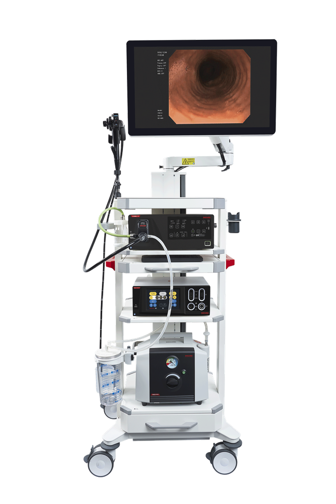
An endoscopy system is a medical imaging platform that lets a clinician see and work inside the body through a natural opening or a small incision, without open surgery. It consists of an endoscope — a long, thin insertion tube carrying illumination, an imaging element, and usually a hollow working channel for passing instruments — connected to a video processor and light source console that turns the captured image into a real-time display. Early endoscopes were purely fiber optic (a bundle of glass fibers relaying the image directly to an eyepiece); virtually all modern clinical systems are video endoscopes, carrying a miniature CCD or CMOS image sensor at the tip that converts light into an electronic signal transmitted up the shaft to the processor. Flexible endoscopes (gastroscopes, colonoscopes, bronchoscopes) bend to follow the body’s natural passages; rigid endoscopes (laparoscopes, arthroscopes) are used through a small surgical port. The FDA regulates the endoscope-and-accessories category as a Class II device under 21 CFR 876.1500.
Everything below unpacks that definition — from imaging-sensor physics, through device architecture and reprocessing requirements, to clinical performance and where the technology is heading.
1. Why endoscopic imaging is a distinct engineering problem
An endoscope has to deliver a diagnostic-quality image through a tube a few millimeters wide, often bent through tight anatomical curves, while simultaneously carrying illumination to a dark body cavity and, frequently, a channel wide enough to pass biopsy forceps, snares, or irrigation fluid — all without the tip overheating, breaking a hermetic seal, or losing image resolution around the bend. That combination of extreme space constraint, mechanical flexibility, and imaging fidelity is why endoscope design, image-sensor placement, and light-source engineering are treated as their own biomedical-device discipline rather than a simple miniature camera.
2. Working principle: illumination, imaging, and channel function
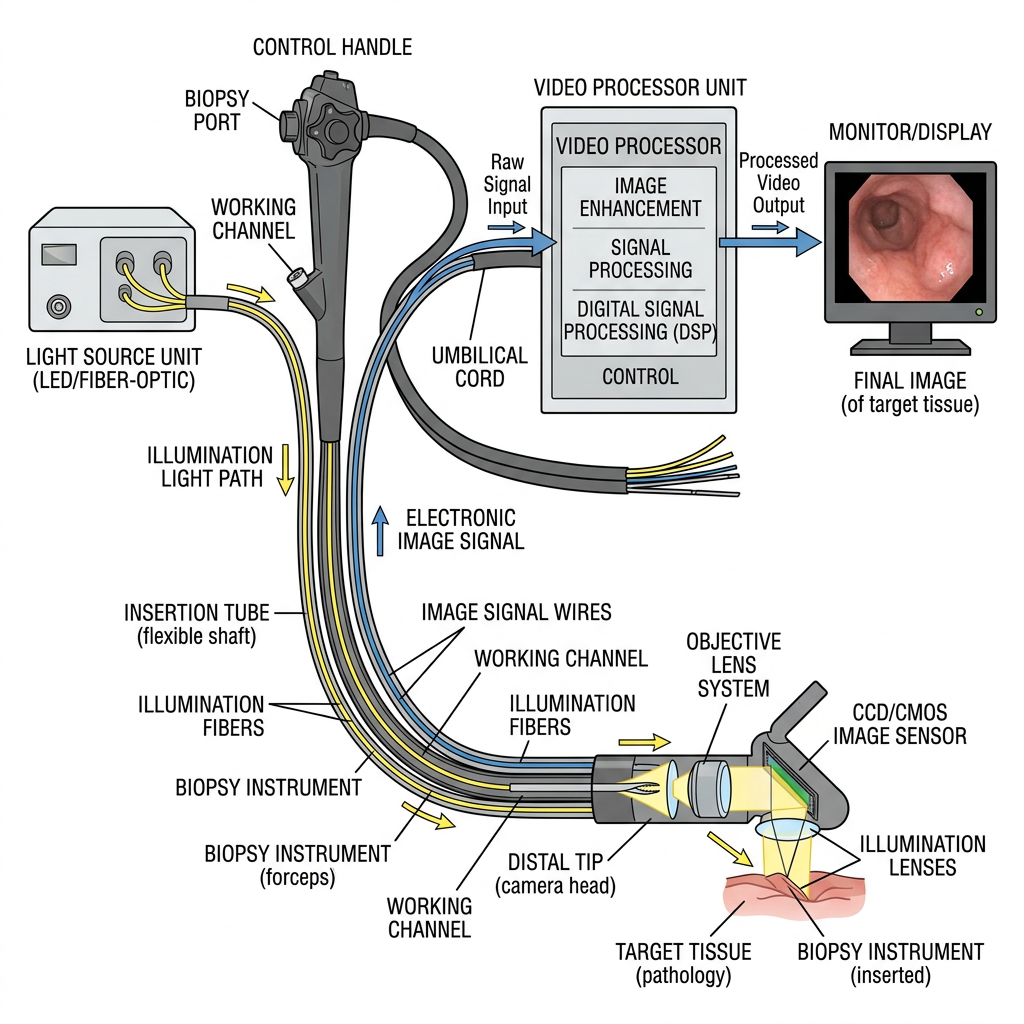
- Illumination. A light source unit — historically halogen or xenon, now predominantly high-output LED in current-generation systems — generates light that travels down the insertion tube, either through dedicated fiber-optic light guides (in both fiber-optic and video endoscopes) or, in some designs, an LED positioned at the distal tip itself.
- Image capture. In a fiber-optic endoscope, a coherent bundle of thousands of individual glass fibers, each preserving its relative position along the bundle, transmits the optical image directly from the objective lens at the tip to an eyepiece or camera adapter at the proximal end — resolution is limited by the number and packing density of individual fibers, and the bundle can show a characteristic honeycomb pattern. In a video endoscope, a miniature CCD (charge-coupled device) or CMOS (complementary metal-oxide-semiconductor) image sensor sits directly behind the objective lens at the distal tip; the sensor converts the optical image into an electronic signal at the point of capture, which is then carried up the shaft as an electrical (rather than optical) signal to the video processor. This chip-at-the-tip design eliminates the fiber bundle’s honeycomb artifact and the cumulative light loss of a long fiber run, and CMOS designs additionally reduce analog noise relative to CCD.
- Video processing. The video processor receives the raw sensor signal, performs image processing (color correction, contrast/edge enhancement, and in current-generation systems, computational image-enhancement modes designed to improve mucosal or vascular contrast), and outputs the processed image to a display in real time.
- Working (instrument) channel. Most flexible diagnostic and therapeutic endoscopes include one or more hollow channels running the length of the insertion tube, used to pass biopsy forceps, snares, clips, or to deliver irrigation and suction — the channel diameter is a key design trade-off against overall insertion-tube diameter and flexibility.
- Steering. The distal tip of a flexible endoscope is articulated by control wires run from angulation knobs on the control body, allowing the operator to steer the tip up/down and left/right to navigate anatomical curves and center the target in view.
3. Device architecture and components
A complete clinical endoscopy system integrates several functional blocks:
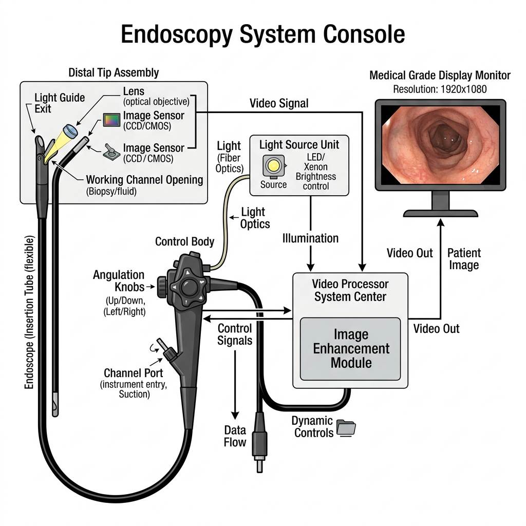
- Insertion tube — the flexible or rigid shaft inserted into the body, containing the imaging path, light guide, working channel(s), and steering control wires (flexible scopes only), engineered to a specific outer diameter and, for flexible scopes, a specific bending radius and insertion-portion length appropriate to the target anatomy.
- Distal tip assembly — houses the objective lens, the CCD/CMOS sensor (video endoscopes) or fiber-bundle terminus (fiber-optic endoscopes), the light-guide exit, and the working-channel opening, all sealed to withstand reprocessing fluids.
- Control body / handle — houses the angulation knobs (flexible endoscopes), instrument-channel port, air/water and suction valves, and remote-control buttons for image capture or processor functions.
- Light source unit — LED or xenon illumination generator, typically combined with the video processor in a single console in current-generation platforms.
- Video processor / system center — receives the sensor signal, applies image processing and enhancement modes, and drives the display; modern platforms increasingly integrate AI-assisted detection and characterization features alongside standard image processing.
- Display monitor — a medical-grade monitor showing the real-time processed image to the operator.
4. Regulatory classification
The FDA regulates endoscopes and accessories under 21 CFR 876.1500, a Class II (moderate risk) device category, reviewed by the Gastroenterology/Urology panel (with related product codes for accessories such as enteroscopes handled under the same regulation number). For reusable flexible endoscopes, a 510(k) submission must include validated reprocessing instructions and reprocessing validation data — a requirement introduced specifically because of the infection-control risk reusable flexible scopes carry.
5. Safety and performance standards
- IEC 60601-2-18 establishes the particular basic safety and essential-performance requirements for endoscopic equipment, sitting alongside the general IEC 60601-1 standard.
- ISO 8600 series (“Endoscopes — Medical endoscopes and endotherapy devices”) covers general requirements (Part 1), optical field-of-view/direction-of-view determination (Part 3), maximum insertion-portion width determination (Part 4), rigid-endoscope optical resolution (Part 5), and vocabulary (Part 6) — the core dimensional and optical-performance standard family for the device class.
- ANSI/AAMI/ISO 17664 and AAMI ST91 govern reprocessing of reusable flexible endoscopes — device manufacturer processing instructions and the facility-level cleaning, high-level disinfection, storage, and quality-control program respectively — the standards underpinning the infection-control side of endoscope use.
6. Clinical use and diagnostic performance
- Gastrointestinal and laparoscopic use. Flexible video endoscopes (gastroscopes, colonoscopes, enteroscopes, bronchoscopes) are used transorally, transanally, or via natural airway access for direct visualization and biopsy/therapeutic intervention within the GI tract and airway; rigid endoscopes (laparoscopes) are inserted through a small abdominal port for minimally invasive surgery.
- Capsule endoscopy as a complementary modality. Rather than a tethered insertion tube, capsule endoscopy uses a swallowed, self-contained imaging capsule to examine regions — particularly the small bowel — that are difficult to reach with a conventional push endoscope. A 2025 review identified five emerging capsule types (steerable, magnetic, robotic, tethered, and hybrid) and reported average per-organ diagnostic accuracy of 96% for the stomach, 98% for the esophagus, and 87% for the colon in current capsule-based examination.
- Magnetically guided capsule endoscopy for upper GI screening. A 2024 study evaluating magnetically guided capsule endoscopy with a detachable string (ds-MCE), using conventional esophagogastroduodenoscopy (OGD) as the reference standard, reported a sensitivity of 97.5% (95% CI 95.5–98.7%) and specificity of 97.8% (95% CI 94.4%–unspecified upper bound) for upper GI lesion detection — supporting capsule-based approaches as a viable, less-invasive screening alternative in appropriate patients.
- AI-assisted image enhancement. Current-generation video processors from major manufacturers now integrate computational image-enhancement modes (e.g., texture and color enhancement) intended to improve mucosal and vascular contrast during real-time examination, part of a broader trend toward AI-assisted detection and characterization support built into the processor rather than added as separate software.
7. QA and testing: what a working system must demonstrate
- Optical field-of-view and direction-of-view verification — per ISO 8600-3, confirming the endoscope’s actual angular field of view and viewing direction match its labeled specification, since a mismatch directly affects lesion detection during a procedure.
- Insertion-portion width verification — per ISO 8600-4, confirming maximum insertion-tube diameter meets the specification relevant to the target anatomical passage (a scope too wide for its intended use is both a usability and a patient-safety issue).
- Optical resolution testing (rigid endoscopes) — per ISO 8600-5, confirming the rigid endoscope’s optical system resolves detail to its rated specification.
- Reprocessing validation — for reusable flexible endoscopes, the manufacturer’s cleaning and high-level-disinfection instructions must be validated to reliably reduce bioburden and biofilm risk in the working channel and other hard-to-reach internal lumens, and this validation data is a required part of the FDA 510(k) submission for the device.
- Leak/seal integrity testing — confirming the insertion tube and distal tip assembly maintain a watertight seal throughout the reprocessing cycle, since a breach allows fluid ingress that can damage internal optics/electronics and compromise sterility between patients.
8. Complications, safety, and failure modes
- Incomplete reprocessing and cross-contamination. The working channel’s narrow, angled internal geometry is difficult to fully clean and disinfect; incomplete reprocessing of reusable flexible endoscopes has been a documented source of patient-to-patient pathogen transmission, which is why reprocessing validation is a mandatory part of the regulatory submission rather than a voluntary quality measure.
- Fiber-bundle image degradation. In fiber-optic endoscopes, individual fiber breakage accumulates over the device’s service life, progressively degrading image resolution and introducing dark spots in the transmitted image — a failure mode largely eliminated by the shift to chip-at-the-tip video endoscopes.
- Insertion-tube mechanical fatigue. Repeated flexing and reprocessing cycles stress the insertion tube’s internal components (control wires, channel liners, electrical/optical conduits); mechanical fatigue can manifest as reduced steering responsiveness or channel leaks well before any visible external damage.
- Light-source thermal risk. High-intensity illumination sources generate heat at the distal tip; excessive exposure duration or a malfunctioning cooling path can pose a localized thermal injury risk to adjacent tissue, which is part of why essential-performance requirements under IEC 60601-2-18 specifically address endoscope thermal safety.
9. Maintenance and troubleshooting
| Symptom | Likely cause | Action |
|---|---|---|
| Dim or uneven illumination | Light-source degradation, fiber breakage in the light guide, or a loose light-guide connector | Inspect light-guide connection; check light-source output; replace scope if internal fiber breakage is confirmed |
| Honeycomb pattern or dark spots in the image | Fiber-bundle breakage (fiber-optic endoscopes) or sensor artifact (video endoscopes) | For fiber-optic scopes, assess breakage extent against manufacturer’s replacement threshold; for video scopes, check sensor and processor cabling/connections |
| Reduced tip steering responsiveness | Control-wire fatigue or damage from repeated flex/reprocessing cycles | Bench-test angulation range against specification; escalate for repair if outside tolerance |
| Fluid ingress / leak detected on leak test | Seal breach in insertion tube or distal tip assembly | Remove from clinical use immediately; do not reprocess or reuse until repaired and re-tested — fluid ingress risks both patient safety and internal component damage |
| Residual bioburden after reprocessing | Incomplete manual cleaning of the working channel, or a reprocessing protocol not matched to the manufacturer’s validated instructions | Re-audit reprocessing protocol against ANSI/AAMI/ISO 17664 and AAMI ST91; verify channel brushing and validated disinfectant contact time |
| Working channel instrument passage resistance | Channel liner damage, debris buildup, or instrument-channel mismatch | Inspect and flush the channel; confirm instrument outer diameter is within the channel’s rated tolerance |
10. Manufacturer landscape
| Manufacturer | Representative system | Notes |
|---|---|---|
| Olympus | EVIS X1™ Video System Center (CV-1500) | Combines video processor and LED light source in one compact console; five-LED illumination array supporting multiple observation modes including Texture and Color Enhancement Imaging (TXI™), with AI-assisted detection features marketed as part of the platform |
| PENTAX Medical | INSPIRA video processor with i20c endoscope series | INSPIRA supports imaging up to 4K resolution and is designed to be multi-connectable with earlier-generation endoscopes; the i20c scope series features an ErgoFeel™ control body and up to 210° tip retroflexion (RetroView™) |
11. Future directions
Capsule-based endoscopy continues to expand beyond simple passive diagnostic imaging toward actively steerable, magnetically guided, and even robotic capsule platforms — a 2025 review catalogued five distinct emerging capsule categories (steerable, magnetic, robotic, tethered, and hybrid) as the field moves toward giving capsule systems some of the targeted maneuverability that conventional tethered endoscopes have always had. In parallel, magnetically guided capsule systems are being validated as genuine screening alternatives for upper GI examination, with recent trial data showing diagnostic accuracy approaching that of conventional tethered endoscopy while avoiding the discomfort and sedation requirements of a standard procedure. On the conventional-endoscope side, AI-assisted real-time image enhancement and lesion-characterization features, now standard on current-generation video processors, are likely to keep expanding as the differentiating capability between manufacturer platforms rather than raw optical resolution alone.
Conclusion
An endoscopy system’s engineering achievement is packing illumination, high-fidelity imaging, and often a working instrument channel into a tube a few millimeters across that must also survive repeated flexing, reprocessing, and years of clinical use without failing at the one moment a clinician needs a clear, safe view inside the body. The shift from fragile fiber-optic bundles to chip-at-the-tip CCD/CMOS video endoscopes, paired with rigorous ISO/IEC dimensional and safety standards and mandatory reprocessing validation, is what has made that combination clinically dependable — and capsule-based and AI-assisted imaging are now extending what the same underlying imaging principle can reach.
FAQ
What’s the difference between a fiber-optic endoscope and a video endoscope? A fiber-optic endoscope transmits the optical image directly through a bundle of glass fibers to an eyepiece; a video endoscope captures the image electronically with a CCD or CMOS sensor at the distal tip and transmits an electronic signal instead — eliminating the fiber bundle’s honeycomb artifact and cumulative light loss, which is why nearly all current clinical systems are video endoscopes.
Is capsule endoscopy meant to replace conventional endoscopy? No — it’s a complementary tool, particularly valuable for reaching small-bowel regions that a tethered scope struggles to access, and recent trial data on magnetically guided capsules shows accuracy approaching conventional endoscopy for upper GI screening specifically, not a wholesale replacement across all indications.
Why does reprocessing validation matter so much for reusable endoscopes? The working channel’s narrow, angled internal geometry is genuinely difficult to fully clean, and incomplete reprocessing has been a documented source of patient-to-patient pathogen transmission — which is why the FDA requires validated reprocessing instructions and validation data as part of the 510(k) submission for reusable flexible endoscopes, not as an optional quality add-on.
What does the FDA device class mean for an endoscope? Class II means moderate risk, requiring a 510(k) premarket submission demonstrating substantial equivalence to a predicate device (including, for reusable flexible scopes, validated reprocessing data) rather than the more extensive premarket approval (PMA) pathway required for higher-risk Class III devices.
References
[1]: Lee CM, et al. “Scanning fiber endoscopy with highly flexible, 1-mm catheterscopes for wide-field, full-color imaging.” Journal of Biophotonics. 2010. PMC3163080.
[2]: Vision Systems Design. “Endoscopes use CMOS image sensors.” https://www.vision-systems.com/home/article/16750278/endoscopes-use-cmos-image-sensors
[3]: U.S. FDA. 21 CFR 876.1500 — Endoscope and accessories; Device Class II; Gastroenterology/Urology review panel. https://www.accessdata.fda.gov/scripts/cdrh/cfdocs/cfpcd/classification.cfm?ID=FDA
[4]: Olympus. “EVIS X1™ Video System Center (CV-1500)” — published product page. https://medical.olympusamerica.com/products/CV-1500
[5]: IEC 60601-2-18:2009, “Medical electrical equipment — Part 2-18: Particular requirements for the basic safety and essential performance of endoscopic equipment.”
[6]: ISO 8600 series, “Endoscopes — Medical endoscopes and endotherapy devices” (Parts 1, 3, 4, 5, 6).
[7]: ANSI/AAMI/ISO 17664:2017, “Processing of health care products — Information to be provided by the medical device manufacturer for the processing of medical devices”; AAMI ST91, “Flexible and semi-rigid endoscope processing in health care facilities.”
[8]: Su CC, Chou CK, Mukundan A, et al. “Capsule Endoscopy: Current Trends, Technological Advancements, and Future Perspectives in Gastrointestinal Diagnostics.” Bioengineering. 2025;12(6):613. doi: 10.3390/bioengineering12060613
[9]: Jiang X, et al. “Diagnostic accuracy of magnetically guided capsule endoscopy with a detachable string for screening of oesophagogastroduodenal lesions.” BMJ. 2024;384:e078581. doi: 10.1136/bmj-2023-078581
[10]: PENTAX Medical. “INSPIRA Video Processor” and “i20c endoscope series” — published product pages. https://www.pentaxmedical.com/us/products/video-processors/inspira ; https://www.pentaxmedical.com/us/products/endoscopes/i20c