
How Does Bone Adapt to Mechanical Load? Wolff’s Law, the Mechanostat, and Stress Shielding
Introduction
A cast removed after eight weeks of immobilization reveals a limb that looks thinner and feels weaker than its counterpart. An astronaut returning from six months in microgravity has measurably less bone mineral in the hip and spine. A tennis player’s dominant-arm bone is denser and thicker than the other arm’s. None of this is coincidence, and none of it is passive wear. Bone is a living tissue that continuously senses the mechanical load it carries and rebuilds itself to match that load.
This behavior is not a minor biological curiosity. It is a governing constraint on how orthopedic implants must be engineered, and a core mechanism within the broader field of biomechanics in biomedical engineering. A metal hip stem or bone plate that is mechanically excellent by classical engineering standards — high stiffness, high strength, low weight — can still fail in the body for a purely biomechanical reason: it changes how load reaches the surrounding bone, and the bone remodels itself in response, sometimes to its own detriment. This article explains the mechanical principle (how bone senses and responds to load), the biological structure that carries it out (the osteocyte network), and the engineering consequence that follows directly from it (stress shielding in orthopedic implants).
Table of Contents
- Wolff’s Law: The Observation Before the Mechanism
- The Mechanostat: A Working Model of Bone Adaptation
- How Bone Senses Load: Osteocytes and Mechanotransduction
- From Biology to Mechanics: Cortical and Trabecular Bone Under Load
- The Engineering Problem: Stress Shielding in Orthopedic Implants
- Engineering Responses to Stress Shielding
- Limitations of the Model Engineers Should Understand
- Engineering Considerations
- Key Takeaways
- References
Wolff’s Law: The Observation Before the Mechanism
In the late nineteenth century, German anatomist and surgeon Julius Wolff observed that the internal trabecular architecture of bone aligns with the directions of mechanical loading it habitually experiences, and that bone shape and density change in measurable ways in response to altered loading. This observation is generally referred to today as Wolff’s Law.
Modern reviews of bone physiology are careful to separate what Wolff actually documented (a correlation between load direction and trabecular orientation) from the mechanistic claims attached to it later.[1] Wolff’s original work predates any understanding of cell biology, molecular signaling, or the tissue-level mechanics needed to explain why the correlation exists. Treated as a historical observation, it holds up well. Treated as a quantitative predictive law, it does not — it says that bone adapts to load, not by how much, under what conditions, or through what cellular pathway. That gap is what the mechanostat model was built to close.
The Mechanostat: A Working Model of Bone Adaptation
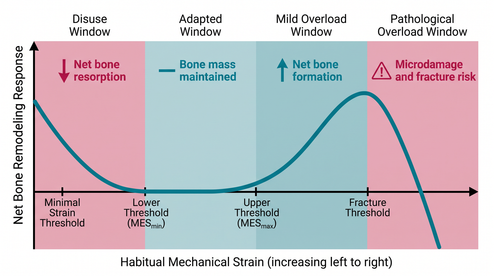
Harold Frost proposed the mechanostat model in 1987 as a more mechanistic and quantitative successor to Wolff’s Law, and updated it in 2003 as evidence accumulated.[2][3] The mechanostat frames bone as a load-sensing feedback system with defined mechanical thresholds, analogous to a thermostat responding to temperature rather than reacting to every fluctuation individually.
The model organizes bone’s response to habitual mechanical strain into zones:
- Disuse window. Strain below a lower threshold signals under-loading. Bone-forming activity falls relative to bone-resorbing activity, and net bone mass decreases. This is the regime behind bed-rest bone loss, spinal-cord-injury osteoporosis, and the bone loss seen in prolonged spaceflight — the same net bone-density loss that a DEXA bone densitometer is designed to quantify clinically.
- Adapted (physiological) window. Habitual strain within this range is treated as adequate; bone mass is maintained near a steady state, with normal remodeling turnover but no net gain or loss.
- Mild overload window. Strain above the adapted range but below the threshold associated with damage triggers net bone formation — the mechanism behind the higher bone density observed in the loaded limbs of athletes.
- Pathological overload window. Strain high enough to risk microdamage triggers a repair-oriented response, and sufficiently repeated or severe overload leads to fatigue microcracks and, eventually, stress fracture.
A 2022 review in Bone Research frames this the same way in contemporary molecular terms: mechanical loading is registered by bone cells, integrated with hormonal and metabolic signals, and converted into a remodeling response that favors formation, maintenance, or resorption depending on the loading history.[4] The thresholds are not universal constants — they vary by skeletal site, age, hormonal status, and loading history — but the framework of upper and lower bounds around a maintained physiological target remains the standard working model for how bone regulates its own mass and architecture.
How Bone Senses Load: Osteocytes and Mechanotransduction
The mechanostat describes the behavior; the osteocyte network is the mechanism that carries it out.
Osteocytes are the most abundant cell type in bone, embedded throughout the mineralized matrix and connected to one another and to the bone surface by long cellular processes running through a network of microscopic channels called the lacunar-canalicular network. This arrangement has led researchers to describe the osteocyte network as bone’s equivalent of a sensory and endocrine organ, distributed through the entire structure rather than located at one site.[5][6]
When bone deforms under load, fluid within the lacunar-canalicular network is forced to flow past the osteocyte processes, generating shear stress on the cell membrane. Osteocytes convert this mechanical signal into a biochemical one — a process called mechanotransduction — through several molecular pathways, most notably the Wnt/β-catenin signaling pathway. A key output of this pathway is the regulation of sclerostin, a protein that normally suppresses bone formation; mechanical loading reduces sclerostin production, releasing that suppression and permitting new bone formation at the loaded site.[7][8] Osteocytes also regulate osteoclast activity — the cells that resorb bone — through signaling molecules such as RANKL and osteoprotegerin, coupling the sensing of insufficient load to an increase in resorption.
This gives the mechanostat a physical substrate: osteocytes measure local strain through fluid flow around their processes, and translate that measurement into the balance between the two remodeling activities — formation by osteoblasts and resorption by osteoclasts — that ultimately determines whether local bone mass rises, falls, or stays the same.
From Biology to Mechanics: Cortical and Trabecular Bone Under Load
Bone tissue exists in two structurally distinct forms that carry load differently. Cortical (compact) bone forms the dense outer shell of long bones and is highly organized, giving it a relatively high stiffness. Trabecular (cancellous) bone forms an open lattice of interconnected struts found at the ends of long bones and inside vertebrae, giving it much lower stiffness and higher porosity. Because the mechanostat responds to local strain rather than to load directly, and strain depends on both the applied load and the local tissue stiffness, the same joint force produces very different strain environments in cortical versus trabecular regions — which is part of why trabecular bone remodels its orientation to align with principal loading directions (the pattern Wolff originally documented) while cortical bone primarily adjusts its thickness and cross-sectional geometry.
This distinction matters directly for implant engineering: an implant placed against cortical bone interacts with a much stiffer, less porous structure than one placed against trabecular bone, and the local strain — and therefore the remodeling signal — the surrounding bone actually experiences depends on how well the implant’s stiffness is matched to the stiffness of the bone it replaces or replaces load-bearing duty for.
The Engineering Problem: Stress Shielding in Orthopedic Implants
This is where mechanobiology becomes an implant design constraint rather than a physiology curiosity.
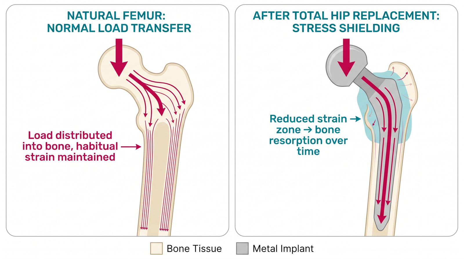
In a healthy femur, mechanical load passes through the bone itself, generating the habitual strain that keeps the mechanostat’s feedback loop in its adapted window. After a total hip replacement, a metal stem is implanted into the femoral canal to transfer load from the joint to the bone. Because common implant alloys — titanium alloys and cobalt-chromium alloys, selected in the first place for their mechanical strength and their biocompatibility — are considerably stiffer than the surrounding bone, a mismatched stem can carry a disproportionate share of the mechanical load itself, rather than transmitting it fully into the adjacent bone. Osteocytes near the implant then register lower strain than before surgery, and the mechanostat responds exactly as it would in an underused limb: net bone resorption in the shielded region. This phenomenon is called stress shielding.
The foundational description of the mechanism comes from Huiskes and colleagues, who applied adaptive bone-remodeling theory to prosthetic-stem design using finite element modeling, showing that stem stiffness and geometry directly predict the pattern of bone loss around a hip implant.[9] Weinans and colleagues followed with a widely cited clinical and analytical account connecting stress shielding directly to the periprosthetic bone resorption observed radiographically around total hip stems, and showed that more flexible stem materials reduce — though do not eliminate — the effect.[10]
The clinical consequence is not cosmetic. Bone lost to stress shielding around a stem is bone no longer available to support future revision surgery if the implant ever needs replacing, and severe periprosthetic bone loss is itself a recognized risk factor for aseptic loosening and periprosthetic fracture. The irony that gives this problem its teaching value: making the implant more mechanically capable in the classical sense — stiffer, stronger — can make the biomechanical outcome worse, because the relevant design target is not the implant’s own strength but the strain it leaves behind in the bone.
Engineering Responses to Stress Shielding
Contemporary implant design addresses stress shielding on several fronts simultaneously, and current biomaterials research is still actively refining all of them:
- Reducing implant stiffness toward bone-matched levels. Design changes such as reduced stem cross-section, proximal-only fixation geometries, and lower-modulus titanium alloys aim to narrow the stiffness mismatch between implant and the biomaterials properties of the bone it replaces, without sacrificing the fatigue strength the stem needs under cyclic gait loading.
- Porous and lattice structures. Additively manufactured porous titanium implants lower the effective bulk stiffness of the implant itself and can be tuned regionally, while also improving bone ingrowth at the implant surface; recent work continues to refine porous titanium surface treatments for both mechanical compatibility and osseointegration.[11]
- Load-sharing geometry over pure material substitution. Because strain — not implant stiffness alone — is what osteocytes respond to, stem geometry that preserves proximal load transfer into bone (rather than shunting load past it to the distal stem tip) can reduce shielding even with a stiffer bulk material.
- Computational prediction before manufacture. Finite element bone-remodeling simulations, following the same adaptive framework Huiskes established, are used as a pre-clinical step for predicting the periprosthetic bone response of a candidate stem design before it is manufactured or tested in patients.[9]
None of these approaches eliminates stress shielding outright; each is a different way of narrowing the mismatch between implant mechanics and the strain environment the surrounding bone’s mechanostat is tuned to expect.
Limitations of the Model Engineers Should Understand
The mechanostat is a useful working framework, not a precise predictive equation. Several qualifications matter for engineering use:
- Strain thresholds vary by skeletal site, age, sex, hormonal status (particularly estrogen and parathyroid hormone signaling), and disease state, so a threshold value measured in one context should not be assumed to transfer directly to another.
- The model describes a systems-level tendency; it does not by itself specify the exact remodeling rate, and computational remodeling models built on it require site-specific calibration against experimental or clinical data before their quantitative predictions can be trusted for a new implant design.
- Stress shielding is a necessary but not sufficient explanation for implant loosening; infection, wear debris osteolysis, malalignment, and initial fixation quality are independent failure pathways that can coexist with it.
Engineering Considerations
For a biomedical engineer, the mechanostat model reframes bone from a passive structural material into an active control system, which changes what “good” implant mechanical design means:
- Design target shifts from implant strength to bone strain. An implant only “succeeds” mechanically if the strain it leaves in the adjacent bone stays inside that bone’s adapted window — an implant can be structurally overbuilt and still bring about a poor biomechanical outcome.
- Stiffness matching is a first-order design variable, not a secondary consideration, in any load-bearing implant that shares mechanical duty with living bone — hip and knee stems, spinal fixation hardware, and dental implants all face a version of the same problem.
- Time matters as much as magnitude. Remodeling is a slow process (months), so post-operative loading progression, immobilization duration, and rehabilitation loading protocols are themselves biomechanical design variables, not purely clinical ones.
- The same feedback loop that causes stress shielding is also the therapeutic target of exercise and pharmacological bone-loss interventions, which is why understanding mechanotransduction has applications well beyond implant design, including osteoporosis management and spaceflight countermeasures.
Key Takeaways
- Wolff’s Law is the nineteenth-century observation that bone architecture aligns with habitual loading direction; the mechanostat (Frost, 1987/2003) is the modern quantitative model of how — bone maintains habitual strain within an adapted window by adjusting formation and resorption.
- Osteocytes, sensing fluid-flow-induced shear stress through the lacunar-canalicular network, are the cellular mechanism carrying out this feedback loop, chiefly through Wnt/β-catenin signaling and sclerostin regulation.
- Stress shielding is the direct engineering consequence of this biology: an implant stiffer than the bone it shares load with reduces local strain below the adapted window, and the mechanostat responds with net bone resorption.
- Reducing stress shielding is fundamentally a strain-matching problem, addressed through stem geometry, reduced or graded stiffness, porous structures, and computational bone-remodeling prediction — not simply “make the implant weaker.”
- The mechanostat is a working framework with site-, age-, and hormone-dependent thresholds, not a fixed universal law; it explains a strong general tendency, not an exact quantitative prediction for every patient.
References
- A 2003 update of bone physiology and Wolff’s Law for clinicians. The Angle Orthodontist, 74(1), 3-15 (2004). https://doi.org/10.1043/0003-3219(2004)074%3C0003:AUOBPA%3E2.0.CO;2
- Frost, H.M. Bone “mass” and the “mechanostat”: a proposal. The Anatomical Record, 219(1), 1-9 (1987). https://doi.org/10.1002/ar.1092190104
- Frost, H.M. Bone’s mechanostat: a 2003 update. The Anatomical Record Part A, 275A(2), 1081-1101 (2003). https://doi.org/10.1002/ar.a.10119
- Chen, X. et al. Mechanical regulation of bone remodeling. Bone Research, 10, 16 (2022). https://doi.org/10.1038/s41413-022-00190-4
- Bonewald, L.F. The amazing osteocyte. Journal of Bone and Mineral Research, 26(2), 229-238 (2011). https://doi.org/10.1002/jbmr.320
- Dallas, S.L., Prideaux, M. & Bonewald, L.F. The osteocyte: an endocrine cell … and more. Endocrine Reviews, 34(5), 658-690 (2013). https://doi.org/10.1210/er.2012-1026
- Bonewald, L.F. & Johnson, M.L. Osteocytes, mechanosensing and Wnt signaling. Bone, 42(4), 606-615 (2008). https://doi.org/10.1016/j.bone.2007.12.224
- Qin, L. et al. Molecular mechanosensors in osteocytes. Bone Research, 8, 23 (2020). https://doi.org/10.1038/s41413-020-0099-y
- Huiskes, R. et al. Adaptive bone-remodeling theory applied to prosthetic-design analysis. Journal of Biomechanics, 20(11-12), 1135-1150 (1987). https://doi.org/10.1016/0021-9290(87)90030-3
- Weinans, H., Huiskes, R. & Grootenboer, H.J. The relationship between stress shielding and bone resorption around total hip stems and the effects of flexible materials. Clinical Orthopaedics and Related Research, (274), 124-134 (1992). https://doi.org/10.1097/00003086-199201000-00014
- A clinically translatable coating-free strategy for 3D-printed porous titanium implants. Bioactive Materials, 2026. https://doi.org/10.1016/j.bioactmat.2026.07.022