How Do Resorbable Implants Disappear? Degradation, Erosion, and the Engineering of Controlled Breakdown
Introduction
Most implants are designed to last. A hip stem, a pacemaker can, a heart valve frame — all are engineered so their material properties change as little as possible over decades in the body. A resorbable implant inverts that requirement completely. A resorbable bone screw, a dissolving suture, a drug-eluting microsphere, or a bioresorbable vascular scaffold is designed to fail — in a controlled, predictable, scheduled way — and then leave nothing behind.
That inversion makes the design problem harder, not easier. The implant must hold its mechanical function for exactly as long as the tissue needs support, then lose that function and clear from the body without producing a harmful local environment on the way out. Getting this wrong in either direction produces a real clinical failure: degrade too fast and the construct loses strength before the tissue can carry load, degrade too slowly and the material behaves like a permanent foreign body that was never intended to stay.
This article explains what actually happens chemically and physically when a resorbable implant breaks down, why “degradation” and “erosion” are two different things that engineers must not conflate, and which design variables genuinely control the timeline.
Table of Contents
- Degradation Is Not Erosion: Two Distinct Processes
- The Chemistry: How Hydrolysis Cuts a Polymer Chain
- Bulk Erosion vs Surface Erosion: The Race Between Water and Chemistry
- Why Strength Disappears Before Mass Does
- The Design Variables That Set the Timeline
- The Acidity Problem: When Degradation Products Cause Trouble
- Resorbable Metals: A Different Mechanism Entirely
- Matching Degradation to Healing: The Real Design Target
- Engineering Considerations
- Key Takeaways
- References
Degradation Is Not Erosion: Two Distinct Processes
These two words are used interchangeably in casual writing, and the confusion causes real design errors. Göpferich’s foundational treatment of the subject draws the distinction precisely:[1]
- Degradation is the chemical process: the scission of polymer chains into shorter fragments, typically by hydrolysis. It happens at the molecular scale and is measured by a drop in molecular weight.
- Erosion is the physical process: the loss of material from the implant as degradation products become soluble enough to diffuse away. It happens at the device scale and is measured by mass loss and dimensional change.
Degradation precedes erosion, and the gap between them is where most of the engineering lives. A polymer implant can lose a large fraction of its molecular weight — and with it, most of its mechanical strength — while still weighing essentially what it did on the day of implantation, because the fragments are not yet small enough to dissolve and leave. An engineer who tracks only mass loss will conclude the implant is intact long after it has stopped being able to carry load.
The Chemistry: How Hydrolysis Cuts a Polymer Chain
The synthetic resorbable polymers used clinically — poly(lactic acid) (PLA), poly(glycolic acid) (PGA), their copolymer poly(lactic-co-glycolic acid) (PLGA), and polycaprolactone (PCL) — are all polyesters. The ester bond linking their repeat units is the designed weak point: it is cleavable by water.
When the implant is placed in the body, water from the surrounding tissue fluid penetrates the polymer and attacks the ester linkages, splitting each bond and producing two shorter chains with new carboxylic acid and hydroxyl end groups. Repeated many times, this chain scission reduces a long, entangled, load-bearing polymer network to progressively shorter fragments, and eventually to the monomer acids themselves — lactic acid and glycolic acid in the case of PLGA — which the body can metabolise and clear through normal pathways.[2][3]
Two features of this chemistry matter for design:
- It is largely passive and water-driven. Unlike many natural biomaterials, degradation of these polyesters does not primarily require enzymes. The dominant mechanism is simple hydrolysis, which is part of why their breakdown behaviour can be engineered relatively predictably in vitro and translated to the in vivo setting.[2]
- It is autocatalytic. The acidic end groups generated by chain scission catalyse further hydrolysis. Degradation, in other words, accelerates itself once it begins — which is why the mass-loss curve of a bulk-eroding polymer is not linear but shows a slow induction phase followed by a much faster collapse.[3]
Bulk Erosion vs Surface Erosion: The Race Between Water and Chemistry
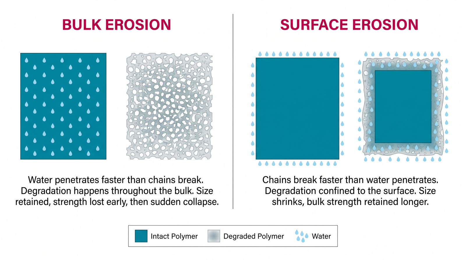
Whether a degradable polymer erodes throughout its volume or only at its outer surface is decided by a competition between two rates: how fast water diffuses into the material, and how fast the chemistry destroys the chains it meets.
- If water penetrates faster than the bonds break, the whole device is wet long before much material is lost. Degradation then proceeds everywhere at once, and the implant hollows out internally while keeping its external dimensions. This is bulk erosion, and it is the behaviour of PLA, PGA, PLGA and PCL.
- If the bonds break faster than water penetrates, the reaction consumes the outermost layer before water can reach the interior. The implant then loses material layer by layer from the outside and shrinks while its core remains intact and undegraded. This is surface erosion, characteristic of much more hydrolytically reactive and hydrophobic polymers such as polyanhydrides.
von Burkersroda, Schedl and Göpferich analysed this competition quantitatively and showed that the erosion mode is not an arbitrary material label but a predictable consequence of the relationship between the polymer’s hydrolysis kinetics, its water diffusivity, and the device’s own dimensions.[4] That last term is important and frequently overlooked: the same polymer can shift its effective erosion behaviour with device size, because a thin fibre and a thick block present very different diffusion distances to water.
The engineering consequences differ sharply:
| Property | Bulk erosion | Surface erosion |
|---|---|---|
| Where degradation occurs | Throughout the whole device | Confined to the outer layer |
| External dimensions over time | Roughly preserved until late collapse | Shrink progressively and predictably |
| Mechanical strength profile | Falls early, well before mass loss | Better retained, since the core stays intact |
| Drug release behaviour | Harder to control; burst release risk at collapse | More nearly linear, near zero-order |
| Local acid build-up | Can concentrate inside the device | Products released directly to the surroundings |
| Representative materials | PLA, PGA, PLGA, PCL | Polyanhydrides, poly(ortho esters) |
Neither mode is universally better. Surface erosion gives more predictable drug release kinetics and better strength retention; bulk-eroding polyesters have far broader clinical history, regulatory familiarity, and processing versatility. The right choice depends on whether the device’s primary job is mechanical support, controlled release, or both.[5]
Why Strength Disappears Before Mass Does
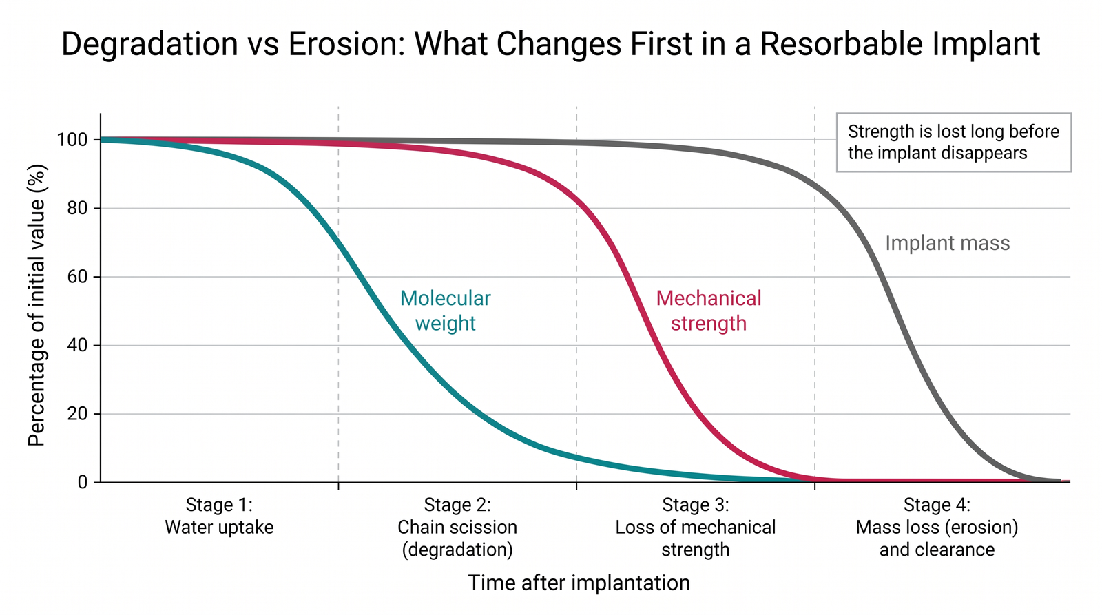
For a bulk-eroding polymer, the property changes follow a consistent sequence, and knowing the order is essential to specifying a resorbable device correctly:
- Water uptake. The device absorbs fluid. Dimensions and mass barely change; the material may soften slightly.
- Chain scission. Molecular weight falls, often steeply. The device still looks and weighs the same.
- Loss of mechanical strength. Once average chain length drops below the level needed for entanglement and load transfer, strength and stiffness fall — still with little mass loss.
- Mass loss and clearance. Fragments finally become soluble, diffuse out, and are metabolised or excreted. Only now does the implant visibly disappear.
The practical rule that falls out of this: a resorbable device’s useful mechanical lifetime is set by stage 3, not by stage 4. A fixation screw described as “resorbing over 18 months” may have lost most of its load-bearing capacity far earlier. Specifying such a device by its total resorption time alone, without a strength-retention profile, is an incomplete specification — and a common source of mismatch between what the engineer intended and what the tissue actually experienced.
The Design Variables That Set the Timeline
Degradation rate is not a fixed property of a material name; it is something the engineer configures. The main levers, drawn from the PLGA literature where they are best characterised:[2][3]
Copolymer composition
In PLGA, the lactide-to-glycolide ratio is the primary control. Glycolic acid units are more hydrophilic and hydrolyse more readily; lactic acid units carry a methyl group that makes the chain more hydrophobic and sterically shields the ester bond. Raising the glycolide fraction generally accelerates degradation; raising the lactide fraction slows it. This single compositional variable lets the same polymer family span device lifetimes from weeks to well over a year.
Molecular weight and end-group chemistry
Higher initial molecular weight means more scission events are needed before fragments become soluble, extending the timeline. End-group chemistry matters too: free carboxylic acid end groups are more hydrophilic and more autocatalytically active than capped ester end groups, so acid-terminated grades degrade faster than end-capped grades of otherwise identical composition.
Crystallinity
Semi-crystalline regions pack polymer chains tightly and exclude water, so they resist hydrolysis far better than disordered amorphous regions. Degradation therefore begins preferentially in the amorphous phase, and a semi-crystalline device can show a two-phase degradation curve — comparatively fast early loss, then a slower tail as the remaining crystalline material breaks down. Crystallinity is set by both composition and processing history, which is why manufacturing route is itself a degradation variable.
Device geometry and porosity
Surface-area-to-volume ratio governs how much material is exposed to water, and wall thickness governs the diffusion distance for both water in and degradation products out. A porous scaffold, a solid screw, and an electrospun fibre mat made from identical polymer stock will not degrade on identical schedules. In thick bulk-eroding devices, trapped acidic products can also accelerate interior degradation relative to the surface.
Sterilisation and storage
Radiation sterilisation can cause chain scission before the device is ever implanted, lowering starting molecular weight and shortening the in-service lifetime. Because hydrolysis is water-driven, moisture exposure during storage does the same. Both belong in the design and specification process, not as afterthoughts.
The Acidity Problem: When Degradation Products Cause Trouble
Polyester degradation releases acid. In a small or well-perfused implant, those products are diluted and cleared without consequence. In a large implant, a thick-walled device, or a poorly vascularised site, they can accumulate faster than the surrounding tissue can buffer them, creating a locally acidic microenvironment that can provoke an inflammatory response and, in some reported clinical cases, sterile fluid collections around resorbable devices.[5]
This links degradation directly to biocompatibility: for a resorbable material, biological safety is not established solely by the parent polymer’s inertness. What matters equally is the concentration, rate and local clearance of the products it generates as it breaks down — which means a resorbable implant’s biological profile depends on its size and site, not only on the material it is made from.
Mitigation strategies used in contemporary design include incorporating basic or buffering fillers such as calcium phosphates, moderating degradation rate so acid generation stays within local clearance capacity, and designing geometry that shortens the diffusion path for degradation products out of the device.[5]
Resorbable Metals: A Different Mechanism Entirely
Where a load-bearing application demands strength and stiffness that polymers cannot deliver, resorbable metals — chiefly magnesium and zinc alloys — offer an alternative. Their disappearance mechanism is corrosion, not hydrolysis: the metal oxidises electrochemically in the body’s chloride-rich aqueous environment, releasing metal ions and, for magnesium, hydrogen gas.
This introduces a distinct set of engineering problems and opportunities:
- Rate control is harder. Corrosion is electrochemical and highly sensitive to alloy composition, microstructure, surface condition and local physiological environment; uncontrolled magnesium corrosion can be too rapid for the healing timeline and can generate hydrogen faster than surrounding tissue absorbs it. Alloy design and protective surface treatments are the principal countermeasures.[6]
- The degradation products are biologically active. Unlike polyester acids, magnesium degradation products participate favourably in the surrounding biology: experimental work reports that they can enhance bone regeneration and exert immunomodulatory and proangiogenic effects, making the degradation process itself part of the therapeutic mechanism rather than merely a tolerated side effect.[7][8]
- Clinical evidence is accumulating. Magnesium-based fixation devices have been evaluated against conventional metal fixation in randomised orthopaedic settings,[9] and bioresorbable magnesium scaffolds have been studied in coronary intervention.[10] Zinc-based systems are at an earlier stage, with reviews framing translation as an open challenge rather than a settled result.[11]
Because resorbable metals are stiffer than the polymers but still less stiff than permanent titanium or cobalt-chromium implants, their mechanical interaction with bone also intersects with stress shielding and bone remodeling — with the added complication that in a resorbable implant, the stiffness mismatch is not constant but changes continuously as the implant corrodes.
Matching Degradation to Healing: The Real Design Target
The central specification for any resorbable device is a rate comparison, not an absolute number: the implant’s loss of mechanical function must track the tissue’s recovery of it.
This is why the same resorbable material can be appropriate in one application and wrong in another. Soft-tissue closure, cancellous bone fixation, cortical bone fixation, and arterial scaffolding all have different healing timescales and different load requirements — and matching them demands different degradation profiles even when the underlying chemistry is the same. Contemporary design work in bioresorbable devices frames this explicitly as designing the degradation profile against the biological timeline rather than selecting a material from a table of properties.[12]
Two persistent difficulties make this harder than it sounds:
- In vitro tests underestimate in vivo complexity. Buffer immersion studies capture hydrolysis but not mechanical loading, cellular activity, local pH variation or perfusion differences, all of which shift the real timeline.
- Healing rates vary between patients. Age, vascularity, comorbidity and mechanical environment all change how fast tissue recovers load capacity, so a degradation profile tuned to an average healing curve will be mismatched at both tails of the patient distribution.
Engineering Considerations
- Specify strength retention, not just resorption time. The clinically relevant parameter is how long the device carries load, which ends in stage 3 — long before the device disappears. A specification that names only total resorption time has not specified the device’s function.
- Treat geometry as a degradation variable. Wall thickness, porosity and surface-area-to-volume ratio change the timeline as much as material choice does, and they can shift a material’s effective erosion behaviour. Never transfer a degradation profile measured on one geometry to a different one.
- Design for the degradation products, not only the material. For polyesters, local acid clearance capacity at the implant site is a real design constraint that scales with device size. For resorbable metals, corrosion rate and hydrogen evolution are the equivalent constraints.
- Include processing and sterilisation in the degradation budget. Thermal history sets crystallinity; radiation sterilisation and moisture exposure lower starting molecular weight. The material as implanted is not the material as supplied.
- Validate under realistic conditions. Static buffer immersion is a screening tool, not a predictor. Loading, flow and a physiologically representative environment are needed before an in vitro degradation profile can support a design claim.
- Materials selection is a systems decision. Choosing between a bulk-eroding polyester, a surface-eroding polymer and a resorbable metal is a trade among strength, degradation predictability, product biology, manufacturability and regulatory precedent — a specific instance of the broader biomaterials selection problem.
Key Takeaways
- Degradation (chemical chain scission) and erosion (physical mass loss) are separate processes; the delay between them is why a resorbable implant can lose its strength while still appearing fully intact.
- The clinically used resorbable polyesters — PLA, PGA, PLGA, PCL — degrade by water-driven hydrolysis of ester bonds, a process that is autocatalytic because its own acidic products accelerate it.
- Bulk erosion versus surface erosion is decided by the competition between water penetration and bond cleavage rates, and depends on device dimensions as well as material chemistry.
- Degradation timelines are engineered, not inherited: copolymer ratio, molecular weight, end-group chemistry, crystallinity, geometry and sterilisation each shift the schedule.
- Degradation products are a design constraint in their own right — acid accumulation for polyesters, corrosion rate and hydrogen evolution for magnesium — and their impact scales with device size and local clearance.
- Resorbable metals degrade by electrochemical corrosion rather than hydrolysis, offer higher strength, and produce degradation products that can actively contribute to healing, though rate control remains the central challenge.
- The correct design target is a match between the implant’s loss of mechanical function and the tissue’s recovery of it, which makes degradation rate an application-specific specification rather than a material property.
References
- Göpferich, A. Mechanisms of polymer degradation and erosion. Biomaterials, 17(2), 103-114 (1996). https://doi.org/10.1016/0142-9612(96)85755-3
- Makadia, H.K. & Siegel, S.J. Poly Lactic-co-Glycolic Acid (PLGA) as biodegradable controlled drug delivery carrier. Polymers, 3(3), 1377-1397 (2011). https://doi.org/10.3390/polym3031377
- Kamaly, N. et al. Degradable controlled-release polymers and polymeric nanoparticles: mechanisms of controlling drug release. Chemical Reviews, 116(4), 2602-2663 (2016). https://doi.org/10.1021/acs.chemrev.5b00346
- von Burkersroda, F., Schedl, L. & Göpferich, A. Why degradable polymers undergo surface erosion or bulk erosion. Biomaterials, 23(21), 4221-4231 (2002). https://doi.org/10.1016/S0142-9612(02)00170-9
- Popescu, B.A. et al. Poly(lactic-co-glycolic acid)-based systems in implantology: advances in biomaterial design and drug delivery. Polymers, 18(9), 1113 (2026). https://doi.org/10.3390/polym18091113
- Hao, M. et al. Hard-soft dual-state coatings regulate degradation rate and biocompatibility of orthopedic magnesium implants. ACS Biomaterials Science & Engineering, 2025. https://doi.org/10.1021/acsbiomaterials.4c01769
- An, Y. et al. Degradation products of magnesium implant synergistically enhance bone regeneration. Bioactive Materials, 2025. https://doi.org/10.1016/j.bioactmat.2024.12.020
- Ben Amara, H. et al. Magnesium implant degradation provides immunomodulatory and proangiogenic effects. Bioactive Materials, 2023. https://doi.org/10.1016/j.bioactmat.2023.02.014
- Saragas, N.P., de Buys, M. & Ferrao, P.N.F. Magnesium bioabsorbable versus titanium screws in hallux valgus surgery: a prospective, randomized study. Foot & Ankle Orthopaedics, 2026. https://doi.org/10.1177/24730114251408829
- Wölbert, S. et al. One-year outcomes of bioresorbable magnesium scaffold implantation in complex coronary lesions. Frontiers in Cardiovascular Medicine, 2026. https://doi.org/10.3389/fcvm.2026.1854686
- Zhao, P. et al. Biodegradable Zn-based implants: progress, challenges, and pathways toward clinical translation. Advanced Science, 2026. https://doi.org/10.1002/advs.76066
- Defining biomaterial-driven design principles for bioabsorbable flow diverters: current state and perspectives. Bioactive Materials, 2026. https://doi.org/10.1016/j.bioactmat.2026.07.045