Why Doesn’t Soft Tissue Behave Like a Spring? Viscoelasticity, Creep, and Stress Relaxation Explained
Introduction
An engineering student’s first mechanical model is a spring. Apply force, get displacement; the ratio is stiffness, and it does not change. Release the force and the spring returns instantly along the same path, giving back all the energy it stored. It is a clean, linear, time-independent, history-free model — and almost every biological soft tissue violates every one of those assumptions.
Pull on a tendon slowly and it feels softer than when you pull quickly. Hold a ligament at fixed length and the force needed to keep it there fades. Rest a load on cartilage and it keeps deforming for minutes. Stretch a strip of arterial wall and release it, and the return path does not retrace the loading path. None of this is measurement error or tissue damage. It is the normal, reproducible mechanical signature of viscoelasticity: material behaviour that combines elastic (solid-like, energy-storing) and viscous (fluid-like, energy-dissipating) responses.
For a biomedical engineer this is not an academic subtlety. It determines what a “stiffness” number from a tissue test actually means, whether a device test protocol produces meaningful results, and how an implant will interact with the tissue around it over time.
Table of Contents
- Elastic, Viscous, and Viscoelastic: The Core Distinction
- The Three Experimental Signatures: Creep, Stress Relaxation, and Hysteresis
- Rate Dependence: Why Loading Speed Changes the Answer
- The J-Shaped Curve: Where Nonlinearity Comes From
- The Structural Origin: What Inside the Tissue Produces This Behaviour
- Modelling Viscoelastic Tissue
- Tissue by Tissue: How the Behaviour Differs
- Why Cells Care: Viscoelasticity as a Biological Signal
- Engineering Considerations
- Key Takeaways
- References
Elastic, Viscous, and Viscoelastic: The Core Distinction
Three idealised behaviours bound the problem:
- A purely elastic solid stores mechanical energy and returns it completely. Stress depends only on current strain, not on how fast or how long it was applied. Its mechanical model is a spring.
- A purely viscous fluid dissipates energy as heat and stores none. Stress depends on strain rate, not on strain itself. Its mechanical model is a dashpot — a piston moving through fluid.
- A viscoelastic material does both. Its stress depends on the current strain, on the rate of loading, and on the loading history that preceded the measurement.
That last clause is what breaks the spring model. In a viscoelastic material, the mechanical response is not a function of the present state alone; the tissue carries a memory of what was done to it. Two identical tendon specimens held at identical strain can be carrying different tension, simply because one was stretched faster or was loaded before.
The Three Experimental Signatures: Creep, Stress Relaxation, and Hysteresis
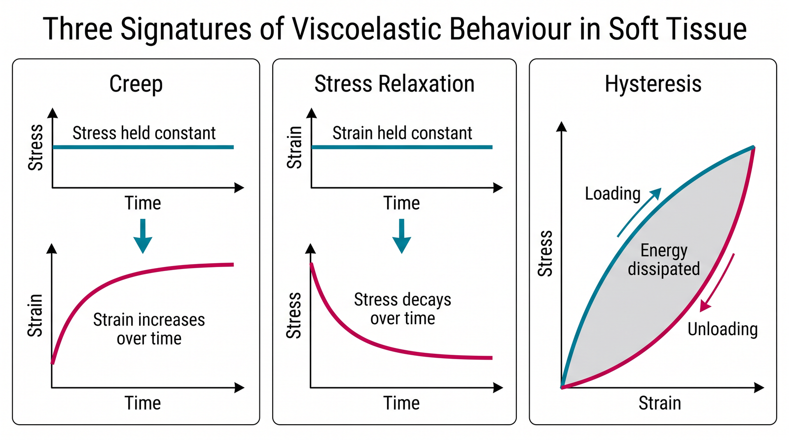
Viscoelasticity shows up in three standard experiments that every tissue-mechanics laboratory runs. Each isolates a different face of the same underlying behaviour.
Creep
Apply a constant stress and hold it. An elastic solid jumps to a fixed strain and stays there. A viscoelastic tissue jumps to an initial strain and then keeps deforming, with strain rising along a decelerating curve toward an equilibrium value.
This is why an intervertebral disc loses height over a day of upright loading, and why prolonged constant pressure on soft tissue produces progressive deformation rather than a single fixed indentation. In cartilage testing, creep response is a standard characterisation method, and the measured properties depend on how long the creep phase is allowed to run before equilibrium is assumed.[1]
Stress relaxation
Apply a fixed strain and hold it. The stress required to maintain that strain falls over time, rapidly at first and then more slowly, toward a non-zero equilibrium plateau.
This is the mirror image of creep, and it is clinically visible: it underlies the gradual loosening of tension in a sutured or tensioned soft-tissue repair and the fading force of orthodontic and tissue-expansion devices. Contemporary ligament studies measure exactly this parameter to characterise and compare tissue bundles.[2]
Hysteresis
Load a tissue and then unload it while recording stress against strain. The unloading curve lies below the loading curve, and the two enclose a loop. The area inside that loop is mechanical energy that was not returned — it was dissipated, mostly as heat.
Hysteresis is why tendon acts as a shock absorber as well as a spring, and why cyclic loading of soft tissue deposits energy in it. It also explains preconditioning: the first several loading cycles of a fresh tissue specimen produce progressively changing curves before the response stabilises into a repeatable loop. Preconditioning is not a laboratory ritual — it is a direct manifestation of viscoelastic history dependence, and results from unpreconditioned first cycles are not comparable with steady-state data.
Rate Dependence: Why Loading Speed Changes the Answer
Because viscous behaviour depends on strain rate, a viscoelastic tissue tested quickly behaves stiffer than the same tissue tested slowly. Load it fast and the viscous component has no time to flow, so the response is dominated by the elastic network; load it slowly and internal fluid movement and molecular rearrangement dissipate part of the input, yielding a more compliant response.
The consequence for measurement is severe: there is no single modulus for a soft tissue. A reported stiffness value is meaningless without the strain rate, the preconditioning protocol, the hydration state, and the temperature at which it was obtained. Rate- and region-dependence has been characterised directly in brain tissue, where mechanical properties vary both with loading rate and with anatomical region.[3][4]
This has direct design consequences. A device that is safe under slow physiological loading may transmit much higher forces during an impact or a rapid manipulation, because the surrounding tissue effectively stiffens at high rates — the mechanism behind many blunt-impact and surgical-tool injury patterns.
The J-Shaped Curve: Where Nonlinearity Comes From
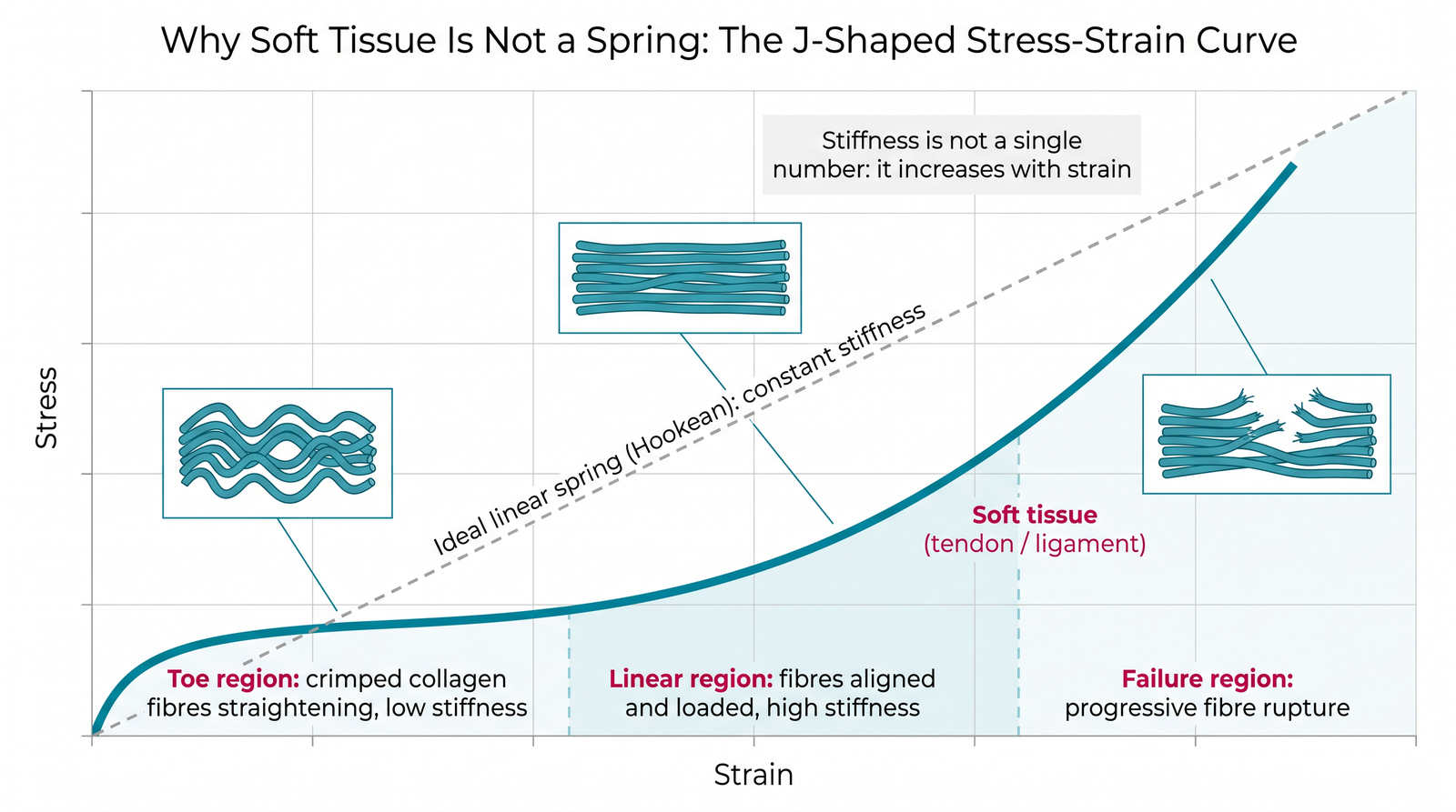
Viscoelasticity is only half of the departure from the spring model. The other half is nonlinear elasticity: even ignoring time effects, stiffness is not constant with strain.
Collagenous soft tissues show a characteristic J-shaped stress-strain curve with three regions:
- Toe region. At low strain, collagen fibres sit in a wavy, crimped configuration. Early deformation mostly straightens this crimp rather than stretching the fibres themselves, so stiffness is low and the tissue is compliant.
- Linear region. Once the crimp is taken up and the fibres align with the load, the stiff collagen molecules themselves carry the load, and stiffness rises sharply to a much higher, roughly constant value.
- Failure region. Beyond a limit, fibres begin to rupture progressively. Stiffness falls, and the tissue fails gradually rather than all at once.
This architecture is functionally important, not incidental. The compliant toe region permits normal joint motion with little resistance, while the stiff linear region resists excessive displacement and protects the joint — a built-in mechanical safety mechanism that a constant-stiffness spring could not provide. Mechanical loading also drives adaptation of tendon structure and function over time, meaning these properties are maintained by use rather than fixed.[5]
The Structural Origin: What Inside the Tissue Produces This Behaviour
Viscoelasticity in soft tissue is not a single mechanism. It emerges from several structural processes acting together:
- Fluid flow through a porous solid matrix. In hydrated tissues, especially articular cartilage, load pressurises interstitial fluid, which then flows slowly through the dense proteoglycan-collagen matrix. This flow dissipates energy and takes time, producing creep and relaxation on long timescales. This mechanism is captured explicitly in biphasic and poroelastic models, and engineered hydrated networks can be designed to reproduce it.[6]
- Collagen fibre reorganisation. Crimp straightening, fibre reorientation toward the load direction, and sliding between fibres and fibrils all take time and dissipate energy.
- Proteoglycan and interstitial matrix behaviour. The ground substance surrounding collagen is highly viscous, and shearing it contributes directly to the viscous response.
- Cellular and active contributions. Living tissue contains cells that can actively generate and modulate tension, adding a component absent from any passive material model.
Because these mechanisms operate on different timescales, real tissue does not relax with a single time constant. It shows a broad spectrum of relaxation times — which is exactly why simple one-spring-one-dashpot models fail to fit tissue data over a wide time range.
Modelling Viscoelastic Tissue
Engineering practice uses a ladder of models, chosen by what the analysis actually needs:
Lumped spring-dashpot models
The Maxwell (spring and dashpot in series), Kelvin-Voigt (parallel), and standard linear solid (three-element) models are built from springs and dashpots. The standard linear solid is the simplest arrangement that reproduces both creep and stress relaxation with a finite equilibrium value, which is why it appears in most textbook treatments. Their strength is analytical transparency; their limitation is that a small number of discrete time constants cannot represent the broad relaxation spectrum of real tissue.
Quasi-linear viscoelasticity (QLV)
Fung’s quasi-linear viscoelastic formulation separates the nonlinear elastic response from the time-dependent relaxation behaviour, treating the relaxation function as independent of strain magnitude.[7] This decomposition made nonlinear tissue data tractable and remains a widely used framework in tissue biomechanics, though its central assumption — strain-independent relaxation — is an approximation that does not hold for every tissue or strain range.
Nonlinear continuum and fibre-reinforced models
For arteries, heart wall, and other fibre-reinforced tissues, anisotropic hyperelastic formulations that explicitly represent collagen fibre families and their orientation distribution are the modern standard, providing physically interpretable parameters tied to real microstructure.[8] These are the constitutive models embedded in finite element solvers written for biological problems, such as the open-source FEBio platform.[9]
Poroelastic and biphasic models
Where fluid flow is the dominant dissipation mechanism — cartilage above all — biphasic models treating the tissue as a porous solid saturated with fluid capture the physics more faithfully than any purely solid viscoelastic model.[1][6]
The right choice is governed by the question, not by sophistication. A suture-pullout analysis may need nothing beyond a relaxation curve; a patient-specific arterial simulation needs an anisotropic fibre-reinforced formulation.
Tissue by Tissue: How the Behaviour Differs
| Tissue | Dominant mechanism | Characteristic behaviour | Typical engineering relevance |
|---|---|---|---|
| Tendon / ligament | Collagen crimp and fibre sliding | Pronounced J-curve, stress relaxation, hysteresis | Graft selection, fixation, suture tensioning[2][5] |
| Articular cartilage | Interstitial fluid flow through porous matrix | Long-timescale creep, load-rate-dependent stiffness | Joint contact mechanics, cartilage repair scaffolds[1][6] |
| Arterial wall | Layered fibre-reinforced structure, residual stress | Anisotropic nonlinear response, hysteresis | Stent and graft design, haemodynamic modelling[8] |
| Brain tissue | Very compliant solid matrix with fluid interaction | Strong rate and region dependence, very low stiffness | Impact and injury modelling, neurosurgical simulation[3][4] |
| Skin | Collagen-elastin network | Nonlinear, anisotropic, direction-dependent | Wound closure, wearable device mechanics |
The common thread: each tissue is viscoelastic, but the mechanism differs, so measurement protocols and models developed for one tissue do not transfer automatically to another.
Why Cells Care: Viscoelasticity as a Biological Signal
For most of the history of tissue mechanics, viscoelasticity was treated as a passive property to be measured. Research over the last decade has shown it is also a biological signal.
Work on engineered matrices with tunable stress relaxation demonstrated that cells respond not only to how stiff their surroundings are, but to how those surroundings relax under the forces the cells themselves apply — with measurable effects on cell spreading, proliferation and differentiation.[10] Subsequent reviews established matrix viscoelasticity as an independent regulator of cellular behaviour across development and disease, distinct from stiffness alone.[11][12][13]
The implication for biomaterials design is substantial: specifying only the elastic modulus of a scaffold or hydrogel leaves out a parameter that cells actively read. This connects tissue viscoelasticity directly to biomaterials design and selection, and parallels the way mechanical signals govern bone adaptation through load-driven remodeling — in both cases, mechanics is an input to biology, not just an output of it.
Measurement technology has followed: magnetic resonance elastography now maps soft-tissue viscoelasticity spatially in vivo, moving these parameters from the test bench toward clinical assessment.[14]
Engineering Considerations
- Never quote tissue stiffness without its protocol. Strain rate, preconditioning history, hydration, temperature and specimen orientation must accompany any modulus value. A number without them is not reproducible and should not be designed against.
- Precondition before measuring, and report it. Steady-state cyclic response is the comparable quantity; first-cycle data reflects the specimen’s prior history, not the tissue’s intrinsic behaviour.
- Test devices at physiologically relevant rates — plural. Because tissue stiffens with rate, a single-rate validation can miss both slow creep failure modes (implant migration, suture loosening) and high-rate failure modes (impact, rapid instrument manipulation).
- Expect long-term relaxation in any tensioned soft-tissue construct. Tension applied at the time of implantation will decrease. Where holding force matters, design for the relaxed equilibrium value, not the initial one.
- Match the model to the question. A lumped spring-dashpot model is adequate for relaxation of a simple construct; fibre-reinforced anisotropic or biphasic formulations are required where fibre orientation or fluid flow dominates.
- Treat viscoelasticity as a design specification for scaffolds, not just a measured property. Since matrix relaxation influences cell behaviour independently of stiffness, a scaffold specification stated only as an elastic modulus is incomplete.
- Mind the boundary between tissue and device. A stiff, nearly elastic implant interfacing with a compliant, time-dependent tissue creates a mechanical mismatch that varies with loading rate — a recurring source of interface stress concentration, micromotion, and long-term loosening. This is one of the central problems in biomechanics as an engineering discipline.
Key Takeaways
- Soft tissue is viscoelastic: its mechanical response depends on strain, on strain rate, and on loading history — so the spring model fails on all three counts.
- The three experimental signatures are creep (strain grows under constant stress), stress relaxation (stress decays under constant strain), and hysteresis (loading and unloading paths differ, dissipating energy).
- Tissue stiffness is not a single number. Any reported modulus is only interpretable alongside the strain rate, preconditioning, hydration and temperature used to obtain it.
- The J-shaped stress-strain curve comes from collagen crimp straightening in the toe region followed by stiff aligned-fibre loading — a built-in mechanism allowing free motion at low load while resisting excessive displacement.
- Different tissues are viscoelastic for different reasons: fluid flow dominates in cartilage, fibre reorganisation in tendon, layered fibre architecture in arteries — so protocols and models do not transfer automatically between them.
- Model selection should follow the engineering question: lumped spring-dashpot models, QLV, anisotropic fibre-reinforced hyperelasticity, and biphasic poroelasticity each answer different classes of problem.
- Viscoelasticity is a biological signal as well as a mechanical property: cells respond to matrix stress relaxation independently of stiffness, which makes it a scaffold design specification rather than just a characterisation result.
References
- Seiferheld, B.E. et al. Confined and unconfined articular cartilage mechanics: effect of creep duration on estimation of material properties. Journal of the Mechanical Behavior of Biomedical Materials, 2025. https://doi.org/10.1016/j.jmbbm.2025.106982
- Ristaniemi, A. et al. Viscoelastic properties of anterolateral and posteromedial bundles of the posterior cruciate ligament. Journal of Biomechanics, 2026. https://doi.org/10.1016/j.jbiomech.2026.113260
- Boiczyk, G.M. et al. Rate- and region-dependent mechanical properties of Göttingen minipig brain tissue in simple shear and unconfined compression. Journal of Biomechanical Engineering, 145(6) (2023). https://doi.org/10.1115/1.4056480
- Jeanpierre, G.M., Rausch, M.K. & Santacruz, S.R. Mechanical properties of fresh rhesus monkey brain tissue. Acta Biomaterialia, 2025. https://doi.org/10.1016/j.actbio.2025.02.049
- Guan, L. et al. Effects of mechanical loading on the structure and function of the Achilles tendon. Journal of Functional Morphology and Kinesiology, 11(3), 273 (2026). https://doi.org/10.3390/jfmk11030273
- Moore, A.C. et al. Fiber reinforced hydrated networks recapitulate the poroelastic mechanics of articular cartilage. Acta Biomaterialia, 2023. https://doi.org/10.1016/j.actbio.2023.06.015
- Fung, Y.C. Biomechanics: Mechanical Properties of Living Tissues, 2nd edition. Springer-Verlag, New York (1993). Origin of the quasi-linear viscoelastic (QLV) formulation for soft tissue.
- Gasser, T.C., Ogden, R.W. & Holzapfel, G.A. Hyperelastic modelling of arterial layers with distributed collagen fibre orientations. Journal of the Royal Society Interface, 3(6), 15-35 (2006). https://doi.org/10.1098/rsif.2005.0073
- Maas, S.A. et al. FEBio: finite elements for biomechanics. Journal of Biomechanical Engineering, 134(1), 011005 (2012). https://doi.org/10.1115/1.4005694
- Chaudhuri, O. et al. Hydrogels with tunable stress relaxation regulate stem cell fate and activity. Nature Materials, 15, 326-334 (2016). https://doi.org/10.1038/nmat4489
- Chaudhuri, O. et al. Effects of extracellular matrix viscoelasticity on cellular behaviour. Nature, 584, 535-546 (2020). https://doi.org/10.1038/s41586-020-2612-2
- Courbot, O. & Elosegui-Artola, A. The role of extracellular matrix viscoelasticity in development and disease. npj Biological Physics and Mechanics, 2025. https://doi.org/10.1038/s44341-025-00014-6
- Eslami, H. & Darvishi, A. Extracellular matrix viscoelasticity: a dynamic regulator of cellular behavior. Annals of Biomedical Engineering, 2025. https://doi.org/10.1007/s10439-025-03767-2
- Zhao, W. et al. Compact tabletop magnetic resonance elastography for mapping soft tissue viscoelasticity. Advanced Science, 2026. https://doi.org/10.1002/advs.75728