How Does a Therapeutic Ultrasound Machine Work? Frequency, Dosage, and Safety Explained — Biomedical Engineering Guide
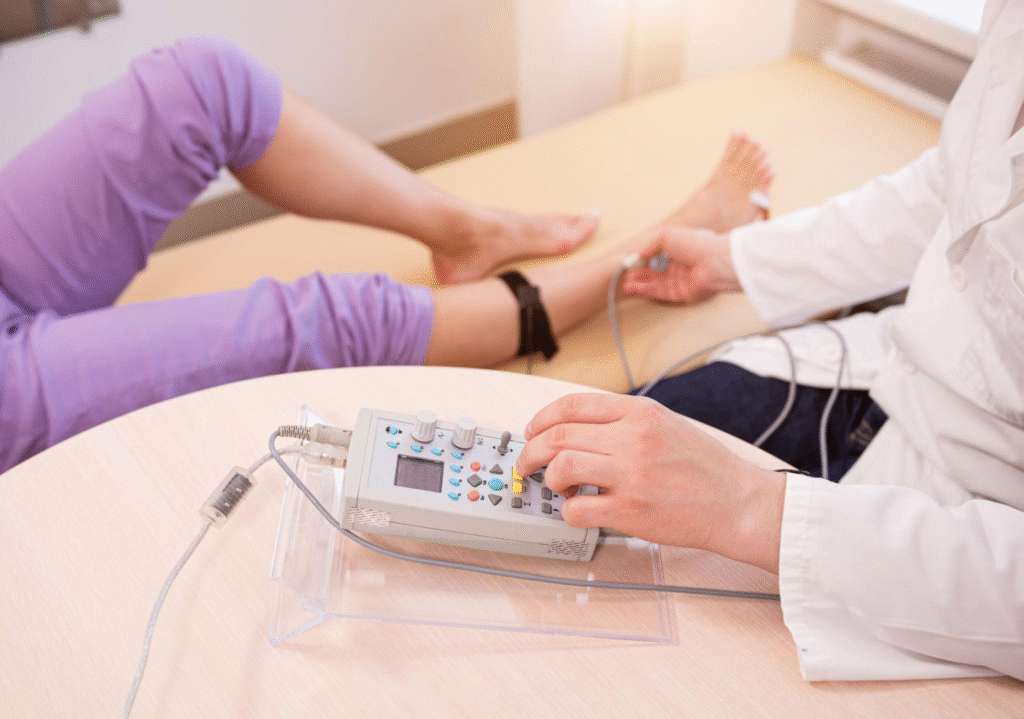
A therapeutic ultrasound machine (also called an ultrasonic diathermy device) is a physiotherapy tool that pushes high-frequency sound waves — well above the range of human hearing — into soft tissue through a handheld applicator, to relieve pain, speed tissue healing, or (at low intensity) stimulate bone repair. A piezoelectric crystal inside the applicator head (usually lead zirconate titanate, PZT) converts an electrical drive signal into mechanical vibration at 1–3.3 MHz; that vibration couples into the patient’s tissue through a water- or gel-based coupling medium. Depending on how the machine is set up, the resulting effect is either thermal — genuine tissue heating for pain relief and increased blood flow, typically using continuous-wave output — or non-thermal/mechanical — cavitation and acoustic streaming at low, pulsed intensities used for tissue-repair stimulation, including bone-fracture healing, with minimal heating. The two modes are configured by the same physical hardware, just different frequency, duty cycle, and intensity settings, and the regulatory classification actually treats them differently: continuous deep-heat ultrasonic diathermy is FDA Class II, while non-thermal therapeutic uses fall under FDA Class III.
Everything below unpacks that definition layer by layer — starting from why the technology exists, through to how it’s built, dosed, tested, and where it’s heading next.
Table of Contents
- 1. Why tissue needs a targeted energy source, not just heat
- 2. Working principle: one physical effect, two very different clinical modes
- 3. Device architecture and components
- 4. Regulatory classification
- 5. Step-by-step operating procedure (thermal/deep-heat mode)
- 6. Materials and power source
- 7. QA and testing: what “delivering the prescribed dose” means numerically
- 8. Complications, safety, and failure modes
- 9. Maintenance and troubleshooting
- 10. Manufacturer landscape
- 11. Choosing between modes and frequencies
- 12. Future directions
- Conclusion
- FAQ
- References
A practical, numbers-first guide for biomedical engineers — physics, device architecture, dosimetry, QA/testing, safety, and where the technology is heading, built on verified 2016–2025 literature.
1. Why tissue needs a targeted energy source, not just heat
Musculoskeletal pain, tendinopathy, and slow-healing fractures often respond to localized heat or mechanical stimulation, but a heating pad or hot pack can only warm the skin and superficial fat — it cannot reliably reach a tendon or joint capsule several centimeters deep without cooking the skin first. Ultrasonic energy solves this differently: sound waves at megahertz frequencies penetrate soft tissue and deposit their energy preferentially at depth, governed by tissue absorption coefficients, so a clinician can target a specific depth by choosing frequency — a genuinely different physical mechanism from surface conduction. The same acoustic-pressure-wave physics, at much lower intensity and in pulsed form, can also mechanically stimulate cells without meaningfully heating them at all, which is what makes low-intensity pulsed ultrasound (LIPUS) usable for stimulating bone-fracture repair without the thermal risk that would come with continuous high-power exposure.2
2. Working principle: one physical effect, two very different clinical modes
The core physics is the piezoelectric effect: an alternating electrical voltage applied across a PZT ceramic disc makes it expand and contract at the drive frequency, launching an acoustic pressure wave into whatever the disc is coupled to. Everything downstream of that — what the ultrasound actually does to tissue — is set by three operator-controlled parameters:
- Frequency — commonly 1 MHz or 3.3 MHz (the “3 MHz” head on many devices) on physiotherapy units. Lower frequency is absorbed more slowly and reaches deeper tissue (on the order of several centimeters), so 1 MHz is used for deeper structures like hip or shoulder musculature; higher frequency is absorbed faster and stays superficial, so 3.3 MHz is used for tendons and tissue close to the skin.
- Duty cycle — the fraction of time the transducer is actually transmitting. Continuous output (100% duty cycle) delivers sustained energy and produces genuine tissue heating (a thermal effect), used for pain relief, muscle relaxation, and increased local blood flow. Pulsed output (commonly 10%, 20%, or 50% duty cycle on physiotherapy devices) delivers the same peak acoustic pressure in short bursts with long off-times between them, so average power stays low and heating is minimal — this is the setup used for non-thermal (mechanical) effects: micro-scale cavitation (oscillating or collapsing gas bubbles at the tissue-fluid interface) and acoustic streaming (fluid movement driven by the sound field), both implicated in accelerating tissue repair.2
- Intensity (measured as spatial-average, temporal-average intensity, ISATA, in W/cm²) — typically 0.5–3 W/cm² for standard physiotherapy applications, but as low as 30 mW/cm² (SATA) for low-intensity pulsed ultrasound (LIPUS) bone-healing devices, which use a 1.5 MHz carrier pulsed at a 1 kHz burst rate with roughly a 20% duty cycle, applied for about 20 minutes per day — an intensity two orders of magnitude below standard physiotherapy dosing, by design, since the mechanism being exploited is mechanotransduction (cells sensing the pressure wave through surface integrins), not heating.3
| Parameter | Thermal / deep-heat mode | Non-thermal / LIPUS bone-healing mode |
|---|---|---|
| Typical frequency | 1 MHz or 3.3 MHz | 1.5 MHz |
| Duty cycle | 100% (continuous) | ~20% pulsed (1 kHz burst rate) |
| Typical intensity (I_SATA) | 0.5–3 W/cm² | 0.03 W/cm² (30 mW/cm²) |
| Session length | 5–10 minutes | ~20 minutes/day |
| Dominant mechanism | Tissue heating | Cavitation / acoustic streaming / mechanotransduction |
| Example device class | Chattanooga Intelect Legend XT | Exogen (Bioventus) |
3. Device architecture and components
- Piezoelectric transducer (applicator head) — a PZT ceramic disc bonded to a metal faceplate; the disc’s diameter and the faceplate together set the applicator’s Effective Radiating Area (ERA) — the actual sound-emitting area, which is always somewhat smaller than the visible faceplate. ERA matters clinically because the same power output produces higher intensity through a smaller ERA.
- Multiple interchangeable applicator heads — clinical systems typically ship 2 cm², 5 cm², and 10 cm² faceplate options so the clinician can match applicator size to the treatment area; on one representative commercial system these correspond to ERAs of 1.8 cm², 4.0 cm², and 8.5 cm² respectively.4
- Drive electronics — generate the RF signal at the selected frequency, gate it to the chosen duty cycle, and regulate output power (0–20 W on the largest head of a representative system, tunable by the clinician).4
- Beam-uniformity control — no real transducer produces a perfectly even acoustic field; the Beam Non-Uniformity Ratio (BNR) — peak intensity divided by the spatial-average intensity — quantifies how “hot-spotty” the beam is. IEC 60601-2-5 caps BNR at 6:1 for physiotherapy equipment; commercial applicators are typically manufactured well under that ceiling (BNR ≤5.0:1 on the representative system above).45
- Coupling medium delivery — the applicator must be acoustically coupled to skin via gel or, for irregular/bony surfaces, an underwater technique; an air gap reflects essentially all the acoustic energy and delivers nothing to tissue.
- Timer and safety interlocks — session timers (accuracy typically within a fraction of a minute) and an output meter that must remain accurate within roughly ±20% of the displayed setting above 10% of maximum output, per representative manufacturer specifications.4
4. Regulatory classification
- United States (FDA) — 21 CFR § 890.5300, “Ultrasonic diathermy,” splits into two very different classifications depending on intended use:
- (a) Deep-heat ultrasonic diathermy — a device generating deep tissue heat for conditions such as pain, muscle spasm, and joint contracture (but not malignancy treatment) is Class II (performance standards; 510(k) premarket notification pathway).
- (b) Ultrasonic diathermy for all other uses — any use not based on generating deep heat (i.e., a non-thermal/mechanical therapeutic claim) is Class III (premarket approval required).1
This means a standard heating-mode physiotherapy ultrasound unit and a device marketed for a non-thermal bioeffect sit in different, non-interchangeable regulatory tiers even though the underlying hardware can be nearly identical. - International standard — IEC 60601-2-5, “Medical electrical equipment — Part 2-5: Particular requirements for the basic safety and essential performance of ultrasonic physiotherapy equipment,” edition 3.0 (2009), specifies the acoustic-output, BNR, ERA labeling, and essential-performance requirements referenced above and is the FDA-recognized consensus standard for this device category.5
- LIPUS bone-healing devices (e.g., Exogen) are cleared under a separate, non-diathermy regulatory pathway specific to their bone-growth-stimulation indication, reflecting that their intended mechanism (mechanotransduction, not heat) is fundamentally different from deep-heat diathermy.
5. Step-by-step operating procedure (thermal/deep-heat mode)
- Confirm the treatment area has no contraindication (see Section 8) and select the applicator size that best matches the treatment area.
- Select frequency based on target depth: 1 MHz for deeper structures (roughly 4–5 cm), 3.3 MHz for superficial structures (roughly 1–2 cm).
- Select duty cycle and intensity per the clinical protocol — typically continuous (100%) at 0.5–3 W/cm² for a thermal/pain-relief session, or pulsed (10–50%) at lower average intensity for an anti-inflammatory/tissue-repair session.
- Apply an adequate layer of acoustic coupling gel to the treatment area (or use an underwater technique for irregular surfaces such as fingers or toes).
- Set the session timer — typically 5–10 minutes per treatment area.
- Continuously move the applicator head in slow, overlapping circular or longitudinal strokes over the treatment area for the full session; holding the head stationary risks a concentrated hot spot due to the beam’s inherent non-uniformity (BNR).
- Monitor the patient for excessive discomfort or a burning sensation — stop immediately if reported, since perceived heat is the clinical feedback signal for a thermal-mode session.
- At session end, remove gel and inspect skin for erythema or any adverse skin reaction before the next session.
6. Materials and power source
- Transducer — lead zirconate titanate (PZT) piezoceramic, chosen for its high electromechanical coupling coefficient at MHz drive frequencies; bonded to a stainless-steel or similar faceplate for durability and acoustic impedance matching.
- Coupling medium — water-based ultrasound gel (never oil-based, which can degrade some faceplate coatings and does not couple as reliably) or a water bath for underwater application.
- Power — mains-powered (AC, e.g., ~100–250 V, 50/60 Hz on a representative clinical system) with regulated low-voltage DC output (e.g., +12 V) driving the RF amplifier stage; clinic-based units are not typically battery-powered given their continuous power draw during treatment, unlike small portable home-use units which run on rechargeable batteries at much lower output power.4
7. QA and testing: what “delivering the prescribed dose” means numerically
- Output power verification — measured with a calibrated radiation-force balance (an acoustic power meter) against the display reading; representative manufacturer tolerance is ±20% of displayed output above 10% of maximum, tested periodically as part of biomedical-engineering preventive maintenance.4
- ERA verification — the applicator’s actual effective radiating area is measured (not just assumed from faceplate diameter) since it directly sets the delivered intensity for a given power setting; ERA is a required label/spec item under IEC 60601-2-5.5
- BNR verification — measured via a scanning acoustic field mapping system; must remain within the 6:1 IEC ceiling (typically well under it, ~5:1, on well-maintained equipment) to avoid unintended tissue hot spots.5
- Timer accuracy — verified against a reference clock; representative tolerance is ±0.2 minute.4
- Clinical follow-up correlate — for a thermal-mode pain/mobility protocol, range-of-motion and pain-scale (e.g., VAS) tracking session-to-session; for LIPUS bone stimulation, serial radiographs assessing fracture-line consolidation are the practical outcome measure, since the device’s job is a biological effect that bench QA alone cannot confirm.
8. Complications, safety, and failure modes
- Contraindicated sites and conditions — ultrasound must not be applied over the eyes, over the heart, over the gravid uterus (especially first trimester), over active malignancy, over areas of acute infection or active DVT/thrombophlebitis, or (per many manufacturer labels) directly over an implanted cardiac device without specific clearance; these are standard physiotherapy contraindications reflected in device labeling and IEC 60601-2-5 warnings.25
- Periosteal burn / bone-interface overheating — because bone absorbs ultrasonic energy far more strongly than soft tissue, holding the applicator stationary over a bony prominence in continuous mode risks a painful periosteal burn; this is why constant applicator motion is a procedural requirement, not just a technique preference.
- Beam non-uniformity hot spots — a BNR above spec (aging transducer, delamination, or manufacturing defect) can create localized tissue heating well above the intended average, even when the displayed output reads normal — a reason for periodic BNR re-verification rather than relying on output-meter accuracy alone.
- Inadequate coupling — an air gap or insufficient gel reflects most of the acoustic energy at the skin surface, meaning the patient receives negligible dose despite an apparently normal-running machine; this is a common, easily overlooked source of “treatment doesn’t seem to work.”
- Cavitation-related bioeffects at excessive intensity — uncontrolled or excessive mechanical-index exposure can theoretically cause unwanted tissue disruption; this is precisely why LIPUS protocols are tightly bounded (30 mW/cm² SATA, ~20% duty cycle) rather than simply “turned down” from standard therapeutic settings.3
9. Maintenance and troubleshooting
| Symptom | Likely cause | Action |
|---|---|---|
| No output despite normal display | Transducer failure, cable fault, or blown fuse | Verify with acoustic output meter; inspect cable/connector; replace fuse per spec (e.g., 4.0 A) |
| Patient reports no sensation of warmth in thermal mode | Inadequate coupling (gel/air gap), applicator not matched to depth, or output below effective threshold | Reapply gel generously; confirm frequency/depth match; verify output with power meter |
| Inconsistent or “patchy” heating sensation | BNR drifted out of spec (transducer wear/delamination) | Bench-test BNR with acoustic field mapping; replace applicator if out of tolerance |
| Output meter reading does not match measured acoustic power | Meter calibration drift | Recalibrate or replace per manufacturer preventive-maintenance schedule |
| Session timer runs long/short | Timer circuit fault | Verify against reference clock; service if outside ±0.2 min tolerance |
| Skin redness/burning reported | Excessive intensity, stationary applicator over bone, or contraindicated site not screened | Stop treatment; review protocol and screening checklist before resuming |
10. Manufacturer landscape
| Manufacturer | Representative product | Technology / notes |
|---|---|---|
| Chattanooga (a DJO/Enovis brand) | Intelect Legend XT | Dual-frequency (1.0/3.3 MHz) clinic system; 2/5/10 cm² applicators with ERA 1.8/4.0/8.5 cm² and BNR ≤5.0:1; pulsed (10/20/50%) or continuous output4 |
| BTL Industries | BTL-4000/5000 series combination therapy units | Ultrasound combined with electrotherapy modalities in a single clinic platform |
| EMS Physio | Various combination and standalone ultrasound therapy units | Clinic-grade physiotherapy ultrasound systems |
| Bioventus | Exogen Ultrasound Bone Healing System | Low-intensity pulsed ultrasound (LIPUS), 1.5 MHz, ~30 mW/cm² SATA, ~20 min/day self-administered for established nonunion fractures6 |
| Mettler Electronics | Sonicator series | Long-standing clinic physiotherapy ultrasound manufacturer |
11. Choosing between modes and frequencies
| Factor | 1 MHz, continuous (deep-heat) | 3.3 MHz, continuous (superficial-heat) | Pulsed, low-intensity (non-thermal/LIPUS) |
|---|---|---|---|
| Target depth | ~4–5 cm | ~1–2 cm | Depends on application; LIPUS targets fracture site directly |
| Primary mechanism | Thermal | Thermal | Mechanotransduction (cavitation/streaming), minimal heat |
| Typical use | Deep muscle, joint capsule, hip/shoulder | Tendon, ligament, superficial scar tissue | Bone nonunion/fracture stimulation |
| Session length | 5–10 min | 5–10 min | ~20 min/day, weeks-long course |
| FDA class (US) | II (deep-heat diathermy) | II (deep-heat diathermy) | Separate bone-growth-stimulator pathway |
12. Future directions
The 2024 update to the field’s major safety/applications review documents continued expansion of therapeutic ultrasound beyond classic physiotherapy — into image-guided focused ultrasound therapies, quality-assurance tooling (tissue-mimicking phantoms, computational dose modeling), and an evolving evidence base correlating specific dosimetry parameters (not just “ultrasound yes/no”) with clinical outcomes.2 For classic physiotherapy-scale devices, the clearest trend is tighter, more standardized dosimetry reporting — ERA, BNR, and duty cycle disclosed and verified per IEC 60601-2-5 — driven by recognition that under-specified older trials made effect sizes hard to compare across studies.
Conclusion
A therapeutic ultrasound machine is, at the hardware level, a single physical mechanism — a piezoelectric transducer converting electrical energy into an acoustic pressure wave — configured two very different ways: continuous, higher-intensity output for genuine tissue heating, and pulsed, very-low-intensity output for non-thermal mechanical stimulation such as bone-fracture healing. The regulatory framework treats these as different device classes precisely because the clinical claim and safety profile differ, even when the underlying transducer technology looks the same. Getting the frequency, duty cycle, and ERA-adjusted intensity right — and verifying them against the IEC 60601-2-5 BNR and output-accuracy limits — is what separates a device that reliably reaches its target and delivers its intended effect from one that merely looks like it’s running.
FAQ
Is therapeutic ultrasound the same machine used for diagnostic imaging? No — diagnostic ultrasound uses very low power and short pulses purely to receive echoes for imaging; therapeutic ultrasound intentionally deposits energy into tissue and uses entirely different power levels and duty cycles.
Why does the therapist keep the applicator moving? To avoid a stationary hot spot — the beam is never perfectly uniform (quantified by the BNR), and bone in particular absorbs ultrasonic energy strongly, so a still applicator over a bony area risks a periosteal burn.
Can therapeutic ultrasound heal a fracture on its own? Low-intensity pulsed ultrasound (LIPUS) devices are cleared specifically as an adjunct for established nonunion fractures, used daily over a period of weeks — they are not a substitute for standard fracture management, but a documented stimulation aid.6
Does a higher frequency mean a stronger treatment? No — higher frequency (e.g., 3.3 MHz vs. 1 MHz) means the energy is absorbed faster and stays more superficial; it is a depth-targeting choice, not an intensity setting.
References
-
U.S. FDA. 21 CFR § 890.5300, “Ultrasonic diathermy.” eCFR / Cornell LII, current edition. ↩
-
Bader KB, Bouchoux G, Holland CK. “Overview of Therapeutic Ultrasound Applications and Safety Considerations: 2024 Update.” J Ultrasound Med. 2025;44(3):381-433. doi: 10.1002/jum.16611 ↩↩↩↩
-
Harrison A, Lin S, Pounder N, Mikuni-Takagaki Y. “Mode & mechanism of low intensity pulsed ultrasound (LIPUS) in fracture repair.” Ultrasonics. 2016;70:45-52. doi: 10.1016/j.ultras.2016.03.016 ↩↩
-
Chattanooga USA. “Intelect Legend US Specifications” (technical datasheet), accessed via MedicalExpo product documentation. ↩↩↩↩↩↩↩↩
-
IEC 60601-2-5:2009, “Medical electrical equipment — Part 2-5: Particular requirements for the basic safety and essential performance of ultrasonic physiotherapy equipment,” edition 3.0. ↩↩↩↩↩
-
Palanisamy P, et al. “Low-Intensity Pulsed Ultrasound Stimulation for Bone Fractures Healing: A Review.” Micromachines. 2021. PMC9290611. ↩↩