
The X-ray machines for Biomedical Engineers: Principles, Components, Types, and Clinical Innovations
The X-ray machine stands as one of the most transformative diagnostic instruments in the history of medicine, forming an indispensable pillar of modern medical imaging and clinical decision-making. For biomedical engineers and healthcare technology professionals, understanding the physics, engineering architecture, and clinical utility of X-ray systems is fundamental to designing, maintaining, and optimizing these devices within complex hospital ecosystems. As a core member of the broader family of radiological devices, the X-ray machine interfaces with virtually every clinical specialty, from emergency trauma to oncology and dental diagnostics. This comprehensive guide explores every dimension of X-ray technology — from the quantum physics of photon generation to the latest detector innovations — providing the technical depth that professionals in biomedical device engineering demand.
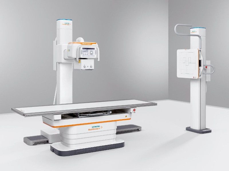
Table of Contents
- What is an X-Ray Machine?
- Why is the X-Ray Machine Used?
- How Does the X-Ray Machine Work in General?
- What Are the Main Components of the X-Ray Machine?
- What Types/Variants of X-Ray Machine Exist?
- What Are the Main Benefits of the X-Ray Machine?
- What Are General Risks or Limitations?
- How is the X-Ray Machine Evolving / Recent Innovations?
- Key Takeaways / Tips for Biomedical Engineers
1. What is an X-Ray Machine?
An X-ray machine is a medical imaging device that produces ionizing electromagnetic radiation in the X-ray spectrum — typically ranging from 20 keV to 150 keV in diagnostic radiology — and projects it through the human body to create images of internal anatomical structures. The differential absorption of X-ray photons by tissues of varying density and atomic composition produces a contrast map that can be captured on film or a digital detector, yielding clinically actionable images. In its most fundamental form, an X-ray machine converts electrical energy into X-ray photon energy through a precisely engineered vacuum tube system, then manages beam delivery, filtration, and detection with a suite of integrated components. As part of the broader landscape of biomedical devices, X-ray systems are classified as Class II or Class III devices under FDA device classification frameworks, reflecting both their diagnostic value and their inherent radiation risk.
A Brief History: From Roentgen’s Discovery to Digital Imaging
The story of the X-ray machine begins on November 8, 1895, when German physicist Wilhelm Conrad Röntgen accidentally discovered a new form of electromagnetic radiation while experimenting with a cathode ray tube in his laboratory at the University of Würzburg. Noticing that a fluorescent screen across the room began to glow despite being shielded from direct cathode rays, Röntgen recognized he had encountered an unknown — and penetrating — form of radiation, which he termed “X-rays” to denote its mysterious nature. Within weeks, he produced the first radiographic image of his wife’s hand, revealing the skeletal structure within and inaugurating a revolution in medical diagnostics. Röntgen was awarded the inaugural Nobel Prize in Physics in 1901 for this discovery.
The subsequent decades saw rapid engineering advancement. Early machines used static anodes and low-power generators, producing images of limited quality with dangerously high radiation doses and no standardized protection protocols. The introduction of the rotating anode tube by physicist Albert Bouwers in the 1920s dramatically increased heat dissipation capacity, allowing for higher tube currents and improved image quality. The mid-20th century brought grid-controlled tubes, automatic exposure control (AEC) systems, and intensifying screens paired with radiographic film, which significantly reduced patient dose. By the 1970s and 1980s, computed radiography (CR) systems using photostimulable phosphor plates began replacing conventional film in many institutions, and by the late 1990s, flat-panel detector technology enabled the full transition to direct and indirect digital radiography (DR). Today, modern X-ray systems are deeply integrated with hospital picture archiving and communication systems (PACS), AI-driven diagnostic tools, and dose management platforms, representing a century of uninterrupted biomedical engineering innovation.
Modern X-Ray Systems and Their Place in Medical Imaging
In the contemporary clinical environment, X-ray machines occupy a central and irreplaceable role within the medical imaging hierarchy. Unlike modalities such as MRI systems or ultrasound machines, X-ray provides unmatched speed, cost-effectiveness, and spatial resolution for osseous and pulmonary pathology assessment. Modern fixed radiography rooms equipped with ceiling-suspended tube assemblies and digital flat-panel detectors can acquire and display diagnostic-quality images in under ten seconds, enabling high-throughput patient workflows in busy emergency departments and outpatient clinics. Furthermore, specialized variants such as fluoroscopy, C-arm systems, dental X-ray units, and mammography systems extend the fundamental X-ray platform to highly specialized clinical domains, each with unique engineering requirements and performance parameters. Understanding how X-ray technology relates to and complements other imaging modalities — including CT scanning and advanced ultrasound technologies — is essential for any biomedical engineer engaged in imaging system selection, procurement, or maintenance.
2. Why is the X-Ray Machine Used?
The enduring clinical prevalence of X-ray machines reflects their extraordinary versatility and the fundamental diagnostic information they provide across virtually every organ system and patient population. With over 3.6 billion X-ray examinations performed globally each year, radiography remains the single most widely utilized medical imaging modality worldwide. For biomedical engineers, appreciating the breadth of clinical applications drives informed decisions about system capability requirements, detector choice, dose optimization strategies, and workflow integration architecture.
Core Clinical Applications Across Medical Specialties
In orthopedics and trauma surgery, plain radiography is the first-line modality for evaluating skeletal injuries, including fractures, dislocations, joint space narrowing in arthritis, and post-surgical hardware assessment. The high spatial resolution of modern flat-panel detectors — reaching pixel pitches below 100 micrometers in some systems — allows visualization of fine cortical bone detail, growth plate abnormalities in pediatric patients, and subtle periosteal reactions indicative of malignancy or infection. In thoracic medicine, chest X-rays remain the cornerstone diagnostic tool for pneumonia, pleural effusion, pneumothorax, cardiomegaly, and pulmonary congestion, providing rapid and cost-effective screening in both acute and ambulatory settings.
Gastroenterology utilizes fluoroscopic X-ray studies — such as barium swallow, barium enema, and upper GI series — to evaluate swallowing disorders, mucosal pathology, and bowel motility. Urology employs intravenous urography and retrograde pyelography for urinary tract assessment. In dentistry, periapical, bitewing, and panoramic X-ray techniques provide irreplaceable anatomical information for caries detection, periodontal bone loss assessment, and implant planning. Meanwhile, mammography — a specialized low-energy X-ray modality — serves as the primary breast cancer screening tool for women over 40, demonstrating the life-saving impact of precisely engineered X-ray systems. For bone density assessment using dual-energy X-ray techniques, the DEXA scanner represents a specialized application of X-ray physics in osteoporosis diagnosis and management.
Role in Emergency Medicine and Critical Care
Few environments demonstrate the clinical indispensability of X-ray machines more vividly than the emergency department and intensive care unit. Portable X-ray units are routinely deployed at the bedside for critically ill patients who cannot be safely transported to a fixed imaging suite, providing immediate assessment of endotracheal tube position, central venous catheter placement, pneumothorax in ventilated patients, and new pulmonary infiltrates in septic patients. Intraoperative C-arm fluoroscopy enables real-time guidance during orthopedic fracture fixation, vascular interventions, and minimally invasive spinal procedures, directly influencing surgical decision-making and outcomes. The speed of acquisition — often under two seconds for a portable AP chest — means that X-ray remains unrivaled in time-critical diagnostic scenarios where delays could directly compromise patient survival.
Oncology and Cancer Screening Applications
X-ray technology plays a multifaceted role in cancer diagnosis and treatment. Chest radiography detects pulmonary nodules that may represent primary lung cancer or metastatic disease, triggering further evaluation with CT. Bone metastases produce characteristic radiographic patterns — lytic, sclerotic, or mixed — that guide staging and treatment planning. In radiation therapy, X-ray-based imaging is fundamental to treatment simulation, portal verification imaging, and on-board kilovoltage imaging systems integrated into linear accelerators. The combination of low cost, rapid acquisition, and wide anatomical coverage makes plain radiography an indispensable first step in many oncological workup pathways, complementing more advanced cross-sectional modalities.
3. How Does the X-Ray Machine Work in General?
The operational principle of an X-ray machine is rooted in atomic physics and electromagnetic theory, involving the controlled generation, shaping, and detection of ionizing photon beams. While the core mechanism — electron acceleration and photon production within a vacuum tube — has remained conceptually consistent since Röntgen’s time, the engineering sophistication surrounding beam generation, control, and detection has advanced enormously. For biomedical engineers, a rigorous understanding of the X-ray generation process is prerequisite to optimizing image quality, managing radiation dose, and diagnosing equipment performance anomalies.
The X-Ray Generation Process
X-ray production begins with thermionic emission: a tungsten filament cathode is resistively heated by a low-voltage filament current (typically 3–5 amperes) to temperatures exceeding 2,200°C, causing electrons to be thermionically emitted from the filament surface into the vacuum of the X-ray tube. The quantity of emitted electrons — and therefore the tube current (mA) — is controlled by the filament heating current, providing the primary mechanism for dose regulation. A high-voltage potential difference, typically ranging from 40 kVp to 150 kVp in diagnostic radiology, is then applied across the tube between the cathode and anode by the high-voltage generator. This accelerates the free electrons through the vacuum gap at velocities approaching a significant fraction of the speed of light, imparting kinetic energies directly proportional to the applied kilovoltage.
Upon striking the tungsten target of the rotating anode, the electrons interact with anode material atoms through two distinct physical mechanisms, both of which produce X-ray photons. Critically, the energy conversion efficiency of this process is extremely low: approximately 99% of the incident electron kinetic energy is converted to heat within the anode, and only approximately 1% is converted into useful X-ray radiation. This thermal burden is the primary engineering challenge in X-ray tube design, necessitating high-speed anode rotation (typically 3,000–10,000 RPM) to distribute heat over a larger focal track area, along with sophisticated anode materials engineering using rhenium-tungsten alloys on graphite or molybdenum substrates for optimized thermal capacity and heat dissipation.
Bremsstrahlung and Characteristic Radiation
The two photon-producing interaction mechanisms within the X-ray tube anode are Bremsstrahlung (braking radiation) and characteristic radiation. Bremsstrahlung is produced when an accelerated electron passes close to the nucleus of a tungsten atom and is deflected by the strong nuclear Coulomb field, losing kinetic energy that is emitted as an X-ray photon. Because the degree of deceleration varies continuously — depending on electron trajectory and nuclear proximity — Bremsstrahlung produces a continuous spectrum of photon energies ranging from zero up to a maximum equivalent to the peak kilovoltage (kVp). The shape of the Bremsstrahlung spectrum is a characteristic ramp that rises from zero at the maximum energy and peaks at approximately one-third of the maximum photon energy.
Characteristic radiation, by contrast, is produced when an accelerated electron ejects an inner-shell orbital electron from a tungsten atom (most commonly from the K-shell), creating a vacancy that is filled by an outer-shell electron cascading inward, with the energy difference emitted as a discrete X-ray photon of characteristic energy. For tungsten, K-characteristic X-rays are produced at approximately 57–69 keV, appearing as discrete spikes superimposed on the Bremsstrahlung continuum in the X-ray spectrum. Characteristic radiation is only produced when the tube voltage exceeds the binding energy of the relevant electron shell — approximately 69.5 keV for tungsten K-shell electrons — and typically contributes approximately 10–30% of the total X-ray output in clinical diagnostic ranges.
Image Formation and X-Ray Attenuation
After exiting the X-ray tube, the X-ray beam is shaped by a collimator and passes through the patient, where photons are differentially attenuated according to the density, atomic number (Z), and thickness of the tissues encountered. The four primary interaction mechanisms responsible for attenuation in the diagnostic energy range are photoelectric absorption, Compton scattering, coherent (Rayleigh) scattering, and pair production — though pair production is negligible below 1.02 MeV and effectively irrelevant in diagnostic radiology. Photoelectric absorption, dominant at lower photon energies and in high-Z materials (bone, contrast agents), produces high image contrast because it varies with the cube of atomic number (Z³) and inversely with the cube of photon energy (E³). Compton scattering, dominant at intermediate to higher diagnostic energies in soft tissue, is largely independent of Z and reduces contrast while contributing to radiation dose and scattered radiation artifacts.
The transmitted photon beam — spatially encoded with the attenuation pattern of the traversed anatomy — is then captured by the detector system (film-screen, CR plate, or DR flat-panel detector), converting the spatial X-ray intensity distribution into a visible image. The resulting radiographic image represents a two-dimensional projection of three-dimensional anatomy, with brightness or density at each image point reflecting the integrated attenuation along the X-ray path through the body. This projectional nature is a fundamental limitation compared to tomographic modalities such as CT scanning, though it is also a source of X-ray’s speed and simplicity advantages.
Beam Control and Image Quality Parameters
Image quality in radiography is governed by four fundamental parameters: contrast, spatial resolution, noise, and artifacts. Contrast is primarily controlled by selecting appropriate kVp — lower kVp increases photoelectric interactions and therefore tissue contrast but raises patient dose, while higher kVp reduces contrast but improves penetration and lowers dose for large body parts. Spatial resolution is determined by focal spot size (smaller focal spots improve resolution but limit heat loading), detector pixel pitch, and geometric magnification factors. Noise — predominantly quantum noise at low exposure levels — is managed through appropriate mAs selection and advanced detector noise reduction algorithms. Beam control hardware, including motorized collimators, added filtration (aluminum, copper), and anti-scatter grids, further shapes the beam quality and optimizes the signal reaching the detector while minimizing scattered radiation artifacts.
4. What Are the Main Components of an X-Ray Machine?
An X-ray system is a precisely integrated assembly of mechanical, electrical, electronic, and optical subsystems, each engineered to contribute to safe, high-quality image acquisition. Biomedical engineers responsible for system procurement, installation, maintenance, or quality assurance must possess a thorough understanding of each major component’s function, design parameters, and failure modes. The following subsections describe the principal engineering components of a modern diagnostic X-ray system.

The X-Ray Tube Assembly
The X-ray tube is the heart of any X-ray machine, and its engineering represents one of the most demanding material science and vacuum technology challenges in medical device manufacturing. A modern diagnostic X-ray tube consists of a glass or metal-ceramic vacuum envelope housing the cathode assembly and rotating anode assembly, mounted within an oil-filled lead-lined housing that provides both electrical insulation and radiation shielding. The cathode assembly typically contains two tungsten filaments of different sizes — a small focal spot (0.3–0.6 mm) for high-resolution imaging and a large focal spot (0.6–1.2 mm) for high-output techniques — mounted within a focusing cup that electrostatically shapes the electron beam onto the anode focal track.
The rotating anode is a disc of rhenium-tungsten alloy (typically 90% W / 10% Re) bonded to a graphite or molybdenum substrate, mounted on a rotor that is magnetically levitated and driven by an external stator coil at 3,000–10,000 RPM. Anode disc diameters range from 80 mm in compact dental tubes to 200 mm in high-capacity angiography tubes. The anode angle (typically 7°–17°) determines the effective focal spot size and the maximum field coverage at the detector, with shallower angles providing smaller effective focal spots at the cost of reduced coverage. Leading tube manufacturers including Siemens Healthineers, Philips, and GE Healthcare each offer proprietary anode and bearing technologies — such as Siemens’ spiral groove bearing (SGB) technology in their Megalix tubes — that significantly extend tube life and thermal capacity beyond conventional ball-bearing designs.

Detection Systems: From Film to Flat-Panel Detectors
The evolution of X-ray detection technology represents one of the most significant engineering advances in medical imaging over the past three decades. Conventional screen-film systems used intensifying screens (gadolinium oxysulfide or calcium tungstate phosphors) to convert X-ray photons to visible light, which then exposed silver halide film. While inexpensive, film systems required chemical processing, offered limited dynamic range, and could not be digitally manipulated or transmitted electronically. Computed radiography (CR) replaced film with reusable photostimulable phosphor (PSP) plates — typically barium fluorohalide compounds — that store a latent image in trapped electron states, which is then read out by a helium-neon laser scanner and converted to a digital image. CR offered significant workflow improvements over film while utilizing existing X-ray equipment, enabling a cost-effective transition to digital imaging in many facilities.
Modern digital radiography (DR) flat-panel detectors represent the current state of the art, providing superior image quality, dramatically faster acquisition, wider dynamic range, and lower effective dose compared to CR or film. Indirect DR detectors use a scintillator layer (typically cesium iodide, CsI, or gadolinium oxysulfide, GOS) to convert X-ray photons to visible light, which is then detected by an amorphous silicon (a-Si) thin-film transistor (TFT) photodiode array. Direct DR detectors — using amorphous selenium (a-Se) as the X-ray photoconductor — convert X-rays directly to electrical charge without an intermediate light conversion step, offering improved spatial resolution for specific applications such as mammography and chest imaging. Detector pixel pitches in modern systems range from 70–200 μm for general radiography to as fine as 50 μm in dedicated high-resolution applications, with active matrix array sizes commonly 35×43 cm or 43×43 cm for full-field chest imaging.
High-Voltage Generator and Control Console
The high-voltage generator is the power electronics core of the X-ray machine, responsible for converting mains AC power into the precisely regulated high-voltage DC supply required by the X-ray tube. Modern generators are high-frequency inverter designs operating at 20–100 kHz, replacing the older single-phase or three-phase transformer-based designs that produced significant voltage ripple and inferior X-ray beam quality. High-frequency generators achieve voltage ripple below 1% (versus 4–13% for three-phase designs and up to 100% for single-phase), resulting in more homogeneous X-ray spectra, improved image contrast, and reduced patient dose. Generator output ranges from 20–30 kW for portable and dental units to 65–100 kW for high-output fixed fluoroscopy and radiography systems. Precise digital control of kVp (±1 kV accuracy), mA (±5% accuracy), and exposure time (down to 1 ms resolution) is essential for reproducible image quality and dose management.
The control console — increasingly implemented as a digital touchscreen workstation integrated with the radiology information system (RIS) and PACS — provides the operator interface for selecting exposure parameters, anatomical program presets, patient data entry, and image review. Advanced consoles from manufacturers such as Siemens Healthineers (MULTIX Impact series), GE Healthcare (Optima series), and Canon Medical (CXDI series) incorporate automatic exposure control (AEC) ionization chambers beneath the detector to automatically terminate exposure when the detector has received sufficient radiation, providing dose-optimized imaging across varied patient body habitus without manual adjustment.
Collimation, Filtration, and Anti-Scatter Grids
Beam management components between the X-ray tube and the patient play a critical role in both image quality and radiation protection. The collimator — a motorized assembly of lead shutters mounted immediately below the X-ray tube housing — restricts the X-ray beam to the area of clinical interest, reducing scatter production and limiting patient dose to non-imaged tissues. Modern light-field/X-ray-field alignment requirements under IEC 60601-1-3 specify that the light field and radiation field must coincide within 2% of the source-to-image distance (SID). Added filtration — aluminum equivalents of 2.5 mm for tubes operated above 70 kVp under FDA 21 CFR 1020.30 requirements — removes low-energy photons that would otherwise contribute only to skin dose without carrying diagnostic information. Compound filters using aluminum and copper combinations (e.g., 1 mm Al + 0.1 mm Cu) are increasingly used for dose reduction in digital systems, exploiting the wide dynamic range of flat-panel detectors to maintain image quality at lower effective doses. Anti-scatter grids — assemblies of parallel lead foils separated by interspace material — are positioned between the patient and detector to absorb obliquely traveling scattered photons, improving contrast by factors of 2–5 at the cost of a 2–5× increase in required exposure (the Bucky factor), and are characterized by their grid ratio (typically 8:1 to 16:1) and line frequency (typically 40–80 lines/cm).
5. What Types and Variants of X-Ray Machines Exist?
The fundamental X-ray platform has been engineered into a diverse family of system variants, each optimized for specific clinical applications, imaging environments, patient populations, and workflow requirements. For biomedical engineers involved in equipment planning, technology assessment, or clinical engineering management, understanding the distinguishing technical characteristics and appropriate use cases of each X-ray system type is essential for making sound procurement and deployment decisions. The following sections describe the principal X-ray machine categories and their defining engineering features.
Fixed Radiography Systems: Conventional to Full Digital
Fixed radiography rooms — the backbone of radiology departments worldwide — have evolved from manual film-based setups to fully digital environments over the past two decades. A modern fixed DR room typically comprises a ceiling-suspended or floor-mounted tube crane assembly, a wall-mounted upright detector stand for chest imaging, a horizontal table-mounted detector for extremity and abdominal studies, and a digital control console. The transition from conventional film to CR and subsequently to DR flat-panel detectors has progressively improved image quality, workflow efficiency, and dose management. Contemporary fixed radiography systems from Siemens Healthineers (MULTIX Impact), GE Healthcare (Definium series), and Philips (DigitalDiagnost series) integrate wireless flat-panel detectors, AI-assisted anatomy recognition, and automatic exposure parameter optimization, representing the pinnacle of current fixed radiography engineering. When assessing fixed radiography alongside more advanced cross-sectional modalities, engineers should consult the CT scanner buying guide to understand how these technologies complement each other in a complete imaging department.
Specialized Variants: Portable, Fluoroscopy, C-Arm, and Dental
Portable X-ray units are self-contained, battery-powered or capacitor-discharge systems mounted on wheeled carriages, enabling bedside imaging in ICUs, operating rooms, emergency bays, and neonatal units. Modern portable DR units incorporate lightweight wireless flat-panel detectors and compact high-frequency generators delivering 25–40 kW output, with direct integration to PACS via Wi-Fi. Fluoroscopy systems provide continuous or pulsed real-time X-ray imaging at 7.5–30 frames per second, enabling dynamic visualization of contrast agent flow, catheter navigation, and joint motion. C-arm fluoroscopy units — named for their characteristic C-shaped arm connecting the X-ray tube to the image intensifier or flat-panel detector — provide flexible isocentric imaging in surgical suites, catheterization laboratories, and pain management clinics, with mobile C-arms offering field portability and fixed surgical C-arms providing high-output performance for complex vascular and orthopedic procedures. Dental X-ray systems, covered in detail in our comprehensive guide to dental imaging systems, range from intraoral periapical units operating at 60–70 kVp to panoramic (OPG) systems and cone beam computed tomography (CBCT) units offering three-dimensional jaw and dentition imaging. The DEXA scanner — while not used for anatomical projection imaging — applies dual-energy X-ray principles to bone mineral density measurement, as described in detail in articles covering DEXA scanner principles.
| Type | Detector | Resolution | Patient Dose | Cost | Workflow Speed |
|---|---|---|---|---|---|
| Conventional Film | Screen-film cassette | High (film-limited) | High (fixed exposure) | Very Low (equipment); ongoing film/chemistry costs | Slow (chemical processing 3–5 min) |
| Computed Radiography (CR) | Photostimulable phosphor (PSP) plate | Moderate (100–200 μm pixel) | Moderate (dose creep risk) | Low-Moderate | Moderate (plate scanning 60–90 sec) |
| Digital Radiography (DR) | Flat-panel (a-Si/CsI or a-Se) | Very High (70–150 μm pixel) | Low (AEC + wide DR) | High | Very Fast (<10 sec to display) |
| Portable X-Ray | Wireless flat-panel DR or CR plate | Moderate-High | Moderate | Moderate | Fast (bedside; <15 sec display with DR) |
| Fluoroscopy | Image intensifier or flat-panel DR | Moderate (real-time priority) | High (continuous exposure) | High | Real-time (7.5–30 fps) |
| C-Arm | Image intensifier or flat-panel DR | Moderate | High (intraoperative use) | High-Very High | Real-time; isocentric positioning |
| Dental X-Ray | Intraoral sensor, PSP plate, or panoramic DR | Very High (intraoral: ~50 μm) | Very Low | Low-Moderate | Fast (intraoral <5 sec; panoramic ~14 sec) |
Each X-ray system type represents a distinct engineering optimization trade-off among image quality, patient dose, workflow speed, system cost, and clinical flexibility. The selection of the appropriate X-ray platform for a given clinical setting requires careful analysis of examination volume, case mix, patient acuity, space constraints, budget parameters, and integration requirements with existing hospital information systems. Biomedical engineers pursuing deeper expertise in medical imaging technology selection and the broader landscape of diagnostic devices may benefit from exploring career development resources in biomedical engineering career pathways and the ethical dimensions of biomedical device innovation, which are central to responsible X-ray system deployment and patient safety stewardship.
6. What Are the Main Benefits of the X-Ray Machine?
Despite being over a century old, the X-ray machine remains one of the most indispensable tools in modern healthcare. Its enduring relevance is not accidental — it reflects a unique combination of clinical utility, operational efficiency, and engineering maturity that few other imaging modalities can match. For biomedical engineers and healthcare technology professionals, understanding these benefits is essential when evaluating procurement decisions, designing clinical workflows, or advising on departmental imaging strategies.
Rapid Diagnostics and Time-Critical Decision Making
One of the most significant advantages of the X-ray machine is its speed. A standard chest radiograph can be acquired in under one second of exposure time, with the entire process — patient positioning, image acquisition, and digital display — typically completed within two to five minutes. This makes X-ray imaging invaluable in emergency and trauma settings, where rapid assessment of fractures, pneumothorax, hemothorax, or foreign body ingestion can directly influence life-saving interventions. In intensive care units, portable bedside X-ray units enable clinicians to monitor endotracheal tube placement, central venous catheter positioning, and pulmonary edema without the logistical burden of transporting critically ill patients. Compared to modalities such as CT scanning or MRI, which require significantly more preparation time, patient cooperation, and infrastructure, plain radiography offers unmatched speed for first-line diagnostic evaluation.
Cost-Effectiveness and Wide Accessibility
X-ray systems represent one of the most cost-effective imaging solutions available in healthcare. Capital costs for a standard digital radiography (DR) system range from approximately $30,000 to $150,000 — a fraction of the investment required for a CT scanner or MRI system. Operational costs, including maintenance, consumables, and staffing, are also substantially lower. This economic advantage makes X-ray imaging accessible not only in well-resourced academic medical centers but also in rural clinics, low-income settings, and field hospitals in resource-limited environments. Portable and battery-operated X-ray units, now widely available from manufacturers such as Carestream, Canon Medical, and Fujifilm, extend this accessibility further, enabling screening programs and diagnostic services in underserved communities worldwide. For healthcare systems managing tight capital budgets, X-ray remains the foundational imaging investment before scaling toward more advanced modalities. Biomedical engineers evaluating departmental imaging strategies should consider exploring resources such as the CT Scanner Buying Guide 2025 to understand cost comparisons across imaging technologies.
Versatility and Broad Clinical Applications
The versatility of X-ray technology is remarkable. A single X-ray system can serve musculoskeletal imaging, chest diagnostics, abdominal surveys, dental applications, fluoroscopic procedures, and mammographic screening — often with appropriate accessory configurations or dedicated system variants. Specialized applications include dual-energy X-ray absorptiometry (DEXA) for bone density assessment, as discussed in detail in our articles on the Bone Densitometer (DEXA Scanner). Dental imaging systems similarly leverage X-ray physics for periapical, bitewing, and panoramic radiography. This broad applicability means that X-ray platforms deliver clinical value across virtually every medical specialty, from orthopedics and pulmonology to oncology and pediatrics. As part of the wider ecosystem of radiological devices, X-ray machines form the backbone of diagnostic imaging infrastructure globally.
Superior Image Quality for Dense Structures
X-ray imaging excels at visualizing structures with high radiodensity, particularly cortical bone, calcified lesions, metallic implants, and consolidated pulmonary tissue. The high inherent contrast between bone and surrounding soft tissue makes plain radiography the gold standard for initial fracture diagnosis, implant surveillance, and assessment of skeletal alignment. In chest imaging, the ability to detect pneumothorax, pleural effusion, cardiomegaly, and pulmonary infiltrates with a single projection remains clinically powerful. While ultrasound and MRI provide superior soft-tissue contrast, no modality rivals the X-ray machine’s ability to render dense anatomical structures with speed, simplicity, and diagnostic reliability.
7. What Are the General Risks and Limitations of the X-Ray Machine?
A thorough understanding of X-ray limitations is as important as appreciating its benefits. Biomedical engineers and clinical technology professionals must be able to communicate these constraints clearly when advising on equipment selection, patient safety protocols, and imaging appropriateness criteria. Awareness of these limitations also drives the innovation cycles that continue to improve X-ray technology.
Ionizing Radiation Exposure and Biological Risk
The most significant limitation of X-ray imaging is its use of ionizing radiation, which carries an inherent biological risk. X-ray photons interact with biological tissues through photoelectric absorption and Compton scattering, depositing energy that can cause DNA strand breaks and, at sufficient cumulative doses, increase the risk of stochastic effects including carcinogenesis. While the effective dose from a single chest radiograph is approximately 0.1 mSv — equivalent to roughly ten days of background radiation — cumulative exposure from repeated imaging, fluoroscopic procedures, or interventional radiology becomes clinically significant. The ALARA (As Low As Reasonably Achievable) principle, enshrined in NCRP Report No. 147 and enforced through regulatory frameworks such as FDA 21 CFR 1020.30, mandates rigorous dose optimization. X-ray is generally contraindicated in pregnant patients, particularly during the first trimester, due to the radiosensitivity of the developing embryo and fetus. These radiation risks stand in direct contrast to non-ionizing modalities such as ultrasound and MRI, which carry no known ionizing radiation hazard. The ethical dimensions of radiation exposure require ongoing vigilance from biomedical engineers who design, procure, and maintain X-ray systems.
Poor Soft-Tissue Contrast and 2D Projection Limitations
Plain radiography produces a two-dimensional projection image that superimposes all anatomical structures along the beam path onto a single plane. This summation effect creates image overlap that can obscure pathology or mimic disease — for example, a pulmonary nodule overlying a rib may be missed, or a vascular shadow may simulate a mediastinal mass. Soft-tissue structures such as the liver, spleen, kidneys, muscles, and neurological tissues are poorly differentiated on plain X-ray due to their similar radiodensity. Contrast agents (barium, iodinated media) are required to enhance visualization of hollow viscera and vascular structures during fluoroscopy, adding procedural complexity and patient risk. For musculoskeletal soft tissue injuries — ligament tears, cartilage damage, meniscal pathology — X-ray provides little diagnostic value, necessitating referral to MRI or ultrasound. These inherent limitations define the boundaries of appropriate X-ray use and underscore the complementary role of modalities such as advanced ultrasound technologies in comprehensive diagnostic imaging strategies.
Operator Dependency and Equipment Maintenance Challenges
Image quality in X-ray radiography is highly dependent on operator skill, including correct patient positioning, appropriate kVp and mAs selection, and accurate collimation. Suboptimal technique can result in under- or overexposed images, rotation artifacts, or unintended anatomy exclusion — all of which reduce diagnostic utility and may necessitate repeat exposures, increasing patient dose. Equipment maintenance is equally critical: aging anode targets, degraded high-voltage generators, misaligned beam collimators, and deteriorating flat-panel detectors can all compromise image quality and dose efficiency. Biomedical engineers are central to establishing preventive maintenance programs, calibration schedules, and quality assurance testing in accordance with standards such as IEC 60601-1-3 and ACR accreditation criteria. Failure to maintain rigorous QA protocols not only degrades clinical performance but also exposes institutions to regulatory and liability risk, as outlined in FDA device classification guidance.
8. How Is the X-Ray Machine Evolving? Recent Innovations and Future Directions
The X-ray machine is far from a static technology. Rapid advances in detector physics, artificial intelligence, and software engineering are transforming what was once a relatively simple projection imaging tool into a sophisticated, data-rich diagnostic platform. These innovations are reshaping dose management, image quality, workflow efficiency, and even the boundaries of anatomical visualization.
Photon-Counting Detectors: The Siemens NAEOTOM Alpha and Beyond
The most transformative detector innovation in recent years is the development of photon-counting computed tomography (PCCT), exemplified by the Siemens Healthineers NAEOTOM Alpha — the world’s first FDA-cleared photon-counting CT scanner, introduced in 2021. Unlike conventional energy-integrating detectors, photon-counting detectors (PCDs) based on cadmium telluride (CdTe) or cadmium zinc telluride (CZT) semiconductors register each individual X-ray photon and measure its energy level directly. This approach eliminates electronic noise contributions, enables intrinsic spectral separation without additional hardware, and produces images with significantly higher spatial resolution — down to 0.2 mm in high-resolution mode. Clinical benefits include sharper delineation of coronary artery stents, inner ear ossicles, and pulmonary micro-nodules, as well as improved material decomposition for iodine and calcium quantification. Dose reduction of 30–50% compared to conventional CT has been demonstrated in early clinical studies. While currently applied primarily in CT, photon-counting detector technology is being explored for next-generation flat-panel radiography systems, potentially delivering spectral imaging capability to routine chest and skeletal X-ray platforms. GE HealthCare and Philips are also actively developing photon-counting solutions for their imaging portfolios.
Artificial Intelligence and Machine Learning in X-Ray Interpretation
Artificial intelligence is rapidly reshaping the diagnostic landscape of radiology, and X-ray imaging is at the forefront of this transformation. Deep learning models trained on large annotated radiographic datasets have demonstrated remarkable capability in detecting pneumonia, tuberculosis, pulmonary nodules, pneumothorax, cardiomegaly, and pleural effusion. Stanford’s CheXNet model, a 121-layer convolutional neural network trained on over 100,000 chest X-rays from the NIH ChestX-ray14 dataset, achieved performance exceeding that of board-certified radiologists on pneumonia detection. Remarkably, AI models have been reported to achieve 61% diagnostic accuracy on complex NEJM case challenges compared to 49% for practicing physicians — a finding with profound implications for diagnostic support systems. FDA-cleared AI tools from companies such as Aidoc, Qure.ai, Zebra Medical Vision, and Annalise.ai are now commercially deployed in clinical radiology departments worldwide, functioning as intelligent triage systems that flag critical findings and prioritize radiologist review queues. For biomedical engineers interested in the expanding role of AI in healthcare, our article on Latest Advances of Artificial Intelligence in Healthcare in 2025 provides a comprehensive overview, and the Practical Roadmap for Biomedical Engineering Students to Learn AI in Healthcare offers actionable guidance for skill development. AI is also driving progress in cancer diagnosis and treatment, with X-ray-based lung cancer screening algorithms demonstrating sensitivity levels approaching low-dose CT in certain populations.
Dose Reduction Technologies and Intelligent Exposure Management
Minimizing patient radiation dose while preserving diagnostic image quality is the central engineering challenge in modern X-ray system design. Advanced automatic exposure control (AEC) systems now incorporate real-time feedback from flat-panel detectors to dynamically modulate kVp and mAs based on patient anatomy and clinical indication. Iterative image reconstruction algorithms adapted from CT — including noise-suppression filters and deep learning-based image enhancement — are being applied to digital radiography to reduce quantum mottle at lower dose levels. Manufacturers including Fujifilm (Dynamic Visualization technology), Agfa (MUSICA processing engine), and Carestream (Directview processing algorithms) have developed sophisticated image processing pipelines that enhance contrast, reduce noise, and optimize spatial frequency response post-acquisition. Pediatric imaging presents a particularly critical application domain for dose reduction, where weight-based and age-adjusted protocols are mandated by campaigns such as the Alliance for Radiation Safety in Pediatric Imaging’s Image Gently initiative. These technological advances ensure that X-ray systems deliver clinically excellent images at doses aligned with international radiation protection guidelines.
Digital Tomosynthesis and Quasi-3D Imaging
Digital tomosynthesis represents a significant step toward three-dimensional information from an X-ray-based platform. By acquiring a limited number of projection images across a short angular sweep (typically 15–60 degrees), reconstruction algorithms generate a series of thin in-focus planes through the anatomy of interest, dramatically reducing the superimposition artifact inherent in conventional radiography. In chest tomosynthesis, studies have demonstrated detection rates for pulmonary nodules approaching those of low-dose CT, at a fraction of the radiation dose. In mammography, digital breast tomosynthesis (DBT) — commercialized by Hologic (Selenia Dimensions) and GE HealthCare (SenoClaire) — has become the new standard of care in many breast imaging programs, reducing recall rates while improving invasive cancer detection. For more on breast imaging technologies, see our detailed guide on Mammography. Musculoskeletal tomosynthesis is also gaining traction for wrist, ankle, and knee imaging, offering improved fracture detection compared to planar radiography without the dose and cost burden of CT.
9. Key Takeaways and Practical Tips for Biomedical Engineers
The X-ray machine occupies a unique and irreplaceable position in the biomedical device ecosystem. For biomedical engineers working in clinical engineering, healthcare technology management, medical device development, or regulatory affairs, a thorough command of X-ray technology fundamentals — combined with awareness of current innovations and compliance requirements — is both professionally valuable and practically essential.
Equipment Selection, Procurement, and Integration
When advising on or managing the procurement of X-ray systems, biomedical engineers should evaluate several critical parameters: detector type and DQE (Detective Quantum Efficiency), spatial resolution (expressed in line pairs per millimeter), system dose efficiency (measured in entrance surface air kerma for standard phantoms), generator power output (kW), and software interoperability with hospital PACS/RIS infrastructure via DICOM and HL7 standards. Compliance with IEC 60601-1 (general safety and essential performance), IEC 60601-1-3 (radiation protection), and ISO 13485 (quality management systems for medical devices) should be verified for all candidate systems. FDA clearance under 21 CFR 1020.30 is mandatory for systems deployed in the United States. Engineers should also assess the vendor’s service infrastructure, availability of spare parts, software update pathways, and AI integration capabilities. Understanding the full spectrum of biomedical device categories enables engineers to contextualize X-ray within a broader imaging strategy that may include CT, MRI, and ultrasound platforms.
Quality Assurance, Preventive Maintenance, and Regulatory Compliance
A rigorous quality assurance program is the cornerstone of safe and effective X-ray operation. Biomedical engineers should implement acceptance testing protocols upon installation, followed by routine performance testing at intervals recommended by AAPM (American Association of Physicists in Medicine) Task Group reports — particularly TG-150 for digital radiography and TG-116 for flat-panel detectors. Key QA parameters include: kVp accuracy and reproducibility, mAs linearity, radiation output consistency, beam collimation accuracy, image uniformity, spatial resolution, low-contrast detectability, and flat-panel detector artifacts (dead pixels, ghosting, lag). Radiation survey measurements should be conducted post-installation and following any system modification, in compliance with NCRP Report No. 147 shielding guidelines. ACR accreditation, while voluntary, provides a structured quality benchmark aligned with best practices. Preventive maintenance schedules should include X-ray tube thermal management, anode rotation bearing inspection, high-voltage cable integrity checks, detector calibration gain and offset corrections, and anti-scatter grid condition assessment. For engineers seeking deeper engagement with regulatory frameworks governing medical devices, our guide on FDA device classification is an essential resource.
Career Development and Expanding Skill Sets
For biomedical engineering professionals, expertise in X-ray technology is a gateway to numerous high-impact career pathways. Clinical engineers managing imaging departments benefit from formal training in medical physics, radiation protection, and PACS administration. Those pursuing roles in medical device development should develop competency in detector physics, X-ray generator design, and regulatory submissions under FDA and CE marking frameworks. The growing integration of AI into X-ray platforms creates particular demand for engineers with combined imaging physics and machine learning skills — a convergence that represents one of the most exciting frontiers in healthcare technology. Our article on Top Career Paths in Biomedical Engineering 2025 outlines how imaging technology expertise translates into diverse professional roles. Engineers early in their careers will also find our Step-by-Step Guide to Becoming a Successful Biomedical Engineer invaluable for structured professional development. Maintaining familiarity with complementary imaging modalities — including the physics of ultrasound acoustic wave interaction and the principles underlying DEXA scanning — broadens an engineer’s clinical utility and positions them as a comprehensive imaging technology resource within their institution. The X-ray machine, in all its evolving sophistication, remains a defining technology of modern medicine — and the engineers who understand it deeply are indispensable partners in delivering safe, effective, and innovative diagnostic care.
References
- Bushberg, J. T., Seibert, J. A., Leidholdt, E. M., & Boone, J. M. (2021). The Essential Physics of Medical Imaging (4th ed.). Wolters Kluwer. Available via academic libraries and medical physics programs.
- U.S. Food and Drug Administration. (2023). Performance Standards for Ionizing Radiation Emitting Products: Diagnostic X-Ray Systems and Their Major Components. 21 CFR Part 1020.30–1020.33. https://www.ecfr.gov/current/title-21/chapter-I/subchapter-J/part-1020
- International Electrotechnical Commission. (2020). IEC 60601-1: Medical Electrical Equipment — Part 1: General Requirements for Basic Safety and Essential Performance (4th ed.). IEC. https://www.iec.ch/publication/2604
- International Electrotechnical Commission. (2020). IEC 60601-1-3: Medical Electrical Equipment — Radiation Protection in Diagnostic X-Ray Equipment. IEC. https://www.iec.ch/publication/22273
- International Organization for Standardization. (2016). ISO 13485: Medical Devices — Quality Management Systems — Requirements for Regulatory Purposes. ISO. https://www.iso.org/standard/59752.html
- National Council on Radiation Protection and Measurements. (2004). NCRP Report No. 147: Structural Shielding Design for Medical X-Ray Imaging Facilities. NCRP. https://www.ncrppublications.org/Reports/147
- Rajpurkar, P., Irvin, J., Ball, R. L., Zhu, K., Yang, B., Mehta, H., … & Lungren, M. P. (2017). CheXNet: Radiologist-level pneumonia detection on chest X-rays with deep learning. arXiv preprint arXiv:1711.05225. https://arxiv.org/abs/1711.05225
- Topol, E. J. (2019). High-performance medicine: The convergence of human and artificial intelligence. Nature Medicine, 25(1), 44–56. https://www.ncbi.nlm.nih.gov/pmc/articles/PMC7015507/
- Siemens Healthineers. (2022). NAEOTOM Alpha: Photon-Counting CT — Technical and Clinical Overview. Siemens Healthineers AG. https://www.siemens-healthineers.com/computed-tomography/photon-counting-ct
- American College of Radiology. (2023). ACR–AAPM Technical Standard for Diagnostic Medical Physics Performance Monitoring of Radiographic and Fluoroscopic Equipment. ACR. https://www.acr.org/Clinical-Resources/Practice-Parameters-and-Technical-Standards
- Lança, L., & Silva, A. (2013). Digital Radiography Detectors — A Technical Overview. SpringerBriefs in Physics. Springer. https://link.springer.com/book/10.1007/978-3-319-01283-5
- Körner, M., Weber, C. H., Wirth, S., Pfeifer, K. J., Reiser, M. F., & Treitl, M. (2007). Advances in digital radiography: Physical principles and system overview. RadioGraphics, 27(3), 675–686. https://pubmed.ncbi.nlm.nih.gov/17495286/
- Vikgren, J., Zachrisson, S., Svalkvist, A., Johnsson, Å. A., Boijsen, M., Flinck, A., … & Bå
th, M. (2008). Comparison of chest tomosynthesis and chest radiography for detection of pulmonary nodules. Radiology, 248(1), 282–288. https://pubmed.ncbi.nlm.nih.gov/18566174/ - Radiology Masterclass. (2023). X-ray Physics and Radiographic Technique. Radiology Masterclass. https://www.radiologymasterclass.co.uk/tutorials/physics
