
SOMATOM On.site: Mobile Computed Tomography Imaging for Critical Care, Emergency Medicine, and Biomedical Engineering Applications
Introduction
Computed tomography (CT) has been one of the most influential medical imaging technologies developed during the past half century. Its ability to generate detailed cross-sectional images of internal anatomy within seconds has made it indispensable for diagnosing acute neurological emergencies, traumatic injuries, vascular diseases, and numerous other conditions. In modern hospitals, CT imaging is often the first diagnostic step when rapid clinical decisions are required.
Despite continuous advances in detector technology, image reconstruction algorithms, and radiation dose optimization, the fundamental workflow of CT imaging has remained remarkably consistent. Patients are transported to the scanner. For stable outpatients and routine examinations, this model works efficiently. However, in critical care environments, the situation becomes far more complex.
A patient in an intensive care unit (ICU) may depend on mechanical ventilation, multiple infusion pumps, invasive hemodynamic monitoring, and continuous neurological assessment. Transporting such a patient to a radiology department is not simply a matter of moving a bed from one room to another. It is a multidisciplinary clinical procedure involving nurses, physicians, respiratory therapists, and transport personnel. During this process, there is always the possibility of physiological instability, interruption of monitoring, accidental disconnection of devices, or delays in treatment.
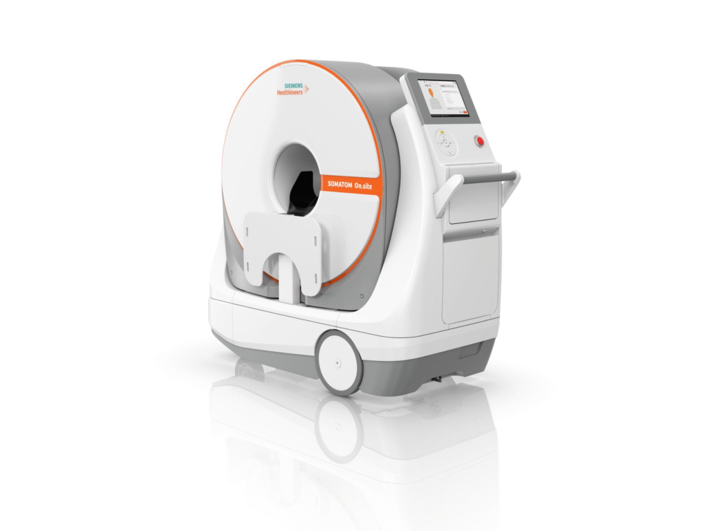
These challenges led to the development of bedside imaging technologies designed to bring diagnostic capabilities closer to the patient. Among the most advanced examples of this concept is SOMATOM On.site, a mobile CT imaging system developed for performing head CT examinations directly within critical care environments.
For biomedical engineers, SOMATOM On.site is particularly interesting because it represents much more than a conventional CT scanner mounted on a mobile platform. It is a complex integration of medical imaging physics, mechanical engineering, radiation safety, embedded computing, hospital workflow design, and healthcare technology management. Understanding how such a system works requires examining not only the clinical need for bedside imaging, but also the engineering principles that make mobile CT technically feasible.
1- What Is SOMATOM On.site?
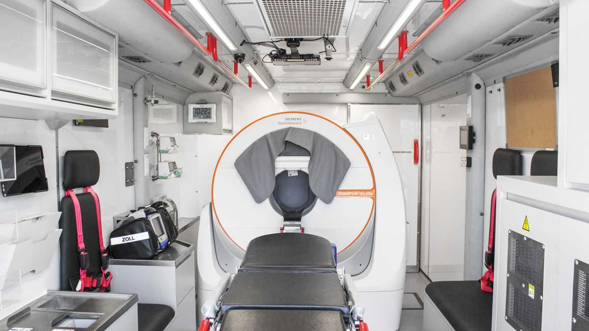
SOMATOM On.site is a mobile head CT imaging system designed to perform computed tomography examinations directly in critical care environments such as ICUs and specialized emergency care settings. Unlike conventional CT scanners that require dedicated installation rooms, the system can be brought to the patient’s location within a healthcare facility.
Purpose and Clinical Role
The primary objective of SOMATOM On.site is to:
- Reduce patient transport requirements
- Accelerate diagnostic decision-making
- Improve access to neuroimaging
- Support critically ill patients who may not tolerate transport
The system is particularly intended for head imaging applications and has been cleared for bedside head CT examinations in ICU environments.
2- Why Mobile CT Imaging Was Developed
The emergence of mobile CT systems reflects a broader trend in healthcare: shifting diagnostic and therapeutic technologies closer to the patient.
Historically, hospitals were designed around centralized departments. Radiology, laboratory medicine, surgery, and intensive care operated as separate units connected through patient transport. While this model remains effective for many clinical activities, it introduces inefficiencies whenever critically ill patients require repeated diagnostic examinations.
Neurocritical care provides a useful example. Patients with traumatic brain injury, intracranial hemorrhage, ischemic stroke, hydrocephalus, or postoperative neurosurgical complications often require serial CT examinations over short periods. Each examination traditionally requires transporting the patient from the ICU to the radiology department and then returning them to the ICU.
From a systems perspective, this workflow creates several challenges. First, transportation consumes clinical resources because multiple healthcare professionals may be required to accompany the patient. Second, transport increases the duration between image request and image acquisition. Third, the movement itself introduces potential clinical risks.
Mobile CT imaging addresses these issues by fundamentally reversing the traditional workflow. Instead of moving the patient to the scanner, the scanner is moved to the patient.
Although this concept appears straightforward, implementing it required solving a series of engineering problems that conventional CT systems rarely encounter.
3- Understanding CT Imaging Fundamentals
To appreciate the design of SOMATOM On.site, it is important to understand the basic principles of CT imaging.
CT scanners produce images using X-rays generated inside a specialized tube. Within the tube, electrons are emitted from a heated cathode and accelerated toward a metal target, typically made from tungsten. When these high-energy electrons collide with the target, their kinetic energy is converted into X-ray photons.
These photons pass through the patient’s body before reaching the detector system located on the opposite side of the gantry. Different tissues absorb different amounts of radiation. Dense structures such as bone attenuate X-rays more strongly than soft tissues, while air-filled spaces attenuate very little radiation.
The detector measures the intensity of the transmitted X-rays from hundreds or thousands of projection angles as the system rotates around the patient. These measurements are then processed mathematically to reconstruct cross-sectional images.
Although this process appears simple conceptually, each stage involves highly sophisticated engineering.
The quality of the final image depends on factors such as:
- X-ray beam stability
- Detector efficiency
- Signal-to-noise ratio
- Mechanical accuracy
- Reconstruction algorithms
- Radiation dose management
A mobile CT system must maintain acceptable performance across all these parameters while operating outside the controlled environment of a conventional CT suite.
4- Detector Technology and Image Formation
The detector system is one of the most important components of any CT scanner because it directly influences image quality.
Modern CT detectors typically use scintillation materials that convert incoming X-ray photons into visible light. This light is then converted into electrical signals by photodiodes. The resulting electrical signals represent the attenuation characteristics of the tissues being imaged.
Detector performance is commonly evaluated through characteristics such as quantum efficiency, dynamic range, spatial resolution, and noise performance.
Quantum efficiency describes how effectively the detector converts incoming X-ray photons into useful signals. Higher efficiency generally improves image quality and may allow lower radiation doses.
Dynamic range determines the detector’s ability to accurately measure both weak and strong signals within the same examination. This is particularly important in head imaging, where dense bone and relatively low-density brain tissue must be visualized simultaneously.
For a mobile CT platform, detector performance becomes even more significant because the system cannot simply compensate for limitations by increasing size, power consumption, or infrastructure requirements. The detector must provide diagnostic-quality performance within a compact and mobile architecture.
5- Engineering Architecture of SOMATOM On.site
Although SOMATOM On.site shares many imaging principles with conventional CT scanners, its architecture has been adapted to support mobility and bedside operation.
At the center of the system is the gantry, which houses the rotating imaging components. The gantry contains the X-ray tube, detector array, collimation systems, and mechanical structures required for precise rotational motion.
The X-ray tube serves as the source of radiation and must maintain stable output during scanning. CT tubes operate under significant thermal stress because much of the electrical energy supplied to the tube is converted into heat rather than useful X-rays. Effective thermal management therefore plays a critical role in maintaining image quality and system reliability.
Opposite the X-ray source lies the detector array, which captures transmitted radiation and converts it into digital signals. These signals are transferred to the data acquisition system, where they are digitized, processed, and prepared for image reconstruction.
Image reconstruction is performed by dedicated computing systems integrated within the scanner architecture. Modern reconstruction algorithms are capable of reducing image noise, minimizing artifacts, and optimizing image quality while controlling radiation dose.
Unlike conventional scanners, however, SOMATOM On.site must also incorporate a mobility platform. This subsystem enables transportation throughout the hospital while maintaining the structural integrity required for accurate imaging.
The mobility platform may appear simple from the outside, but it represents one of the most challenging aspects of the system’s design.
6- Mechanical Stability and Mobility
Conventional CT scanners benefit from permanent installation within dedicated imaging suites. Once installed, the geometric relationship between the X-ray source, detector array, and patient positioning system remains essentially fixed.
Mobile CT systems cannot rely on this advantage.
Every time the scanner is moved, it experiences vibration, acceleration, deceleration, and mechanical loading. Even extremely small changes in alignment can affect image quality because CT reconstruction algorithms assume precise geometric relationships among system components.
For this reason, maintaining mechanical stability is one of the most important engineering challenges associated with mobile CT.
The design must ensure that repeated transportation does not compromise calibration accuracy. Structural rigidity, vibration control, alignment monitoring, and calibration procedures therefore become essential elements of the system architecture.
From an engineering standpoint, mobility is not merely an added feature. It fundamentally changes the design requirements of the scanner.
7- Radiation Safety in Bedside Imaging
Radiation protection is another area where mobile CT systems differ significantly from conventional installations.
Traditional CT scanners operate within specially designed imaging rooms equipped with structural shielding. Walls, doors, and observation windows are engineered to limit radiation exposure to healthcare personnel and nearby patients.
A mobile CT system must function in environments that were not originally designed for CT imaging. Consequently, radiation management becomes a larger responsibility of the device itself.
Radiation safety strategies involve controlling the geometry of the X-ray beam, minimizing unnecessary scatter radiation, and implementing operational procedures that protect nearby staff during examinations.
For biomedical engineers responsible for technology assessment and radiation safety programs, understanding these principles is essential when evaluating mobile imaging systems.
8- Clinical Applications
The primary clinical role of SOMATOM On.site is bedside neuroimaging.
One of its most important applications involves monitoring patients with intracranial hemorrhage. Rapid detection of hemorrhage progression can significantly influence treatment decisions and patient outcomes.
The system is also valuable for evaluating ischemic stroke, particularly when repeated imaging is required to monitor disease evolution or therapeutic response.
In neurocritical care units, bedside CT imaging can facilitate routine neurological assessment without exposing patients to the risks associated with transportation.
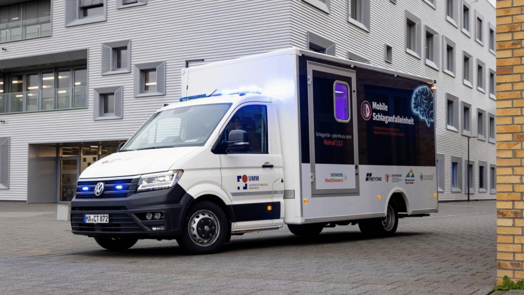
Additional applications include postoperative neurosurgical imaging, emergency neurological assessment, and selected situations in military or disaster medicine where conventional imaging infrastructure may be difficult to access.
9- The Biomedical Engineer’s Perspective
For biomedical engineers, SOMATOM On.site should be viewed not only as an imaging device but also as a healthcare technology system that must be integrated into a complex clinical environment.
The evaluation process begins long before the scanner is used clinically. During technology assessment, engineers must determine whether mobile CT imaging addresses a genuine clinical need and whether the expected benefits justify the investment.
Following installation, acceptance testing verifies that the system performs according to manufacturer specifications and regulatory requirements. Engineers assess image quality, radiation output, safety systems, network connectivity, and interoperability with hospital information systems.
Throughout the operational life of the system, preventive maintenance and quality assurance activities help ensure continued reliability. These activities may include calibration verification, image quality testing, mechanical inspection, and software maintenance.
Because mobile CT systems operate differently from conventional scanners, biomedical engineers must also consider workflow integration, transportation procedures, and environmental factors that may influence long-term performance.
Conclusion
SOMATOM On.site represents a significant development in the evolution of medical imaging because it challenges one of the longest-standing assumptions in radiology: that patients must be transported to the scanner.
Its importance extends beyond mobility alone. The system demonstrates how advances in imaging technology, mechanical design, computing, and healthcare workflow engineering can be combined to create new models of diagnostic care.
For biomedical engineers, SOMATOM On.site provides an excellent case study in multidisciplinary healthcare technology design. It illustrates how solving a clinical problem often requires integrating expertise from medical imaging physics, mechanical engineering, radiation safety, software systems, and clinical operations.
As healthcare continues to move toward faster, safer, and more patient-centered models of care, mobile imaging systems are likely to play an increasingly important role. Understanding the principles behind these technologies will therefore become an essential part of biomedical engineering practice in the years ahead.
References
- McCollough, C. H., Leng, S., Yu, L., & Fletcher, J. G. (2015).
Computed tomography dose optimization and imaging physics principles. Radiologic Clinics of North America, 53(3), 501–515.
https://doi.org/10.1016/j.rcl.2015.02.004 - Hsieh, J. (2009).
Computed Tomography: Principles, Design, Artifacts, and Recent Advances (2nd ed.). SPIE Press.
https://www.spiedigitallibrary.org/ebooks/PM/Computed-Tomography - Bushberg, J. T., Seibert, J. A., Leidholdt, E. M., & Boone, J. M. (2011).
The Essential Physics of Medical Imaging (3rd ed.). Lippincott Williams & Wilkins.
https://shop.lww.com/The-Essential-Physics-of-Medical-Imaging/p/9781451108947
- Kalender, W. A. (2011).
Computed Tomography: Fundamentals, System Technology, Image Quality, Applications (3rd ed.). Publicis.
https://link.springer.com/book/10.1007/978-3-540-74614-5 - Willemink, M. J., & Noël, P. B. (2019).
The evolution of image reconstruction for CT—from filtered back projection to artificial intelligence. European Radiology, 29, 2185–2195.
https://doi.org/10.1007/s00330-018-5810-7
- Rubino, J. G., et al. (2019).
Mobile CT in the intensive care unit: feasibility and clinical impact. Critical Care Medicine, 47(12), e1000–e1006.
https://doi.org/10.1097/CCM.0000000000004021 - Takahashi, S., et al. (2020).
Bedside CT imaging in neurocritical care: reducing transport-related risks. Neurocritical Care, 32(2), 456–463.
https://doi.org/10.1007/s12028-019-00852-1 - PubMed Central (PMC).
Intrahospital transport of critically ill patients: risks and safety considerations.
https://www.ncbi.nlm.nih.gov/pmc/articles/PMC5603353/
- International Electrotechnical Commission (IEC).
IEC 60601-1: Medical electrical equipment – General requirements for basic safety and essential performance.
https://www.iec.ch - International Organization for Standardization (ISO).
ISO 14971: Medical devices — Application of risk management to medical devices.
https://www.iso.org/standard/72704.html - ISO.
ISO 13485: Quality management systems for medical devices.
https://www.iso.org/standard/59752.html
- International Commission on Radiological Protection (ICRP).
The 2007 Recommendations of the ICRP (Publication 103).
https://www.icrp.org/publication.asp?id=ICRP%20Publication%20103 - International Atomic Energy Agency (IAEA).
Radiation Protection in Diagnostic and Interventional Radiology.
https://www.iaea.org/topics/radiation-protection
- Association for the Advancement of Medical Instrumentation (AAMI).
Medical equipment management programs.
https://www.aami.org - ECRI Institute.
Healthcare Technology Management Guidelines and Device Evaluation Reports.
https://www.ecri.org
Siemens Healthineers.
SOMATOM On.site – Mobile CT System Overview.
https://www.siemens-healthineers.com/computed-tomography/mobile-ct/somatom-onsite
