
What Is an Implantable Cardioverter-Defibrillator (ICD)? Architecture, Detection Algorithms, and Biomedical Engineering Guide
An implantable cardioverter-defibrillator (ICD) is a battery-powered active implantable device that continuously monitors heart rhythm and automatically delivers therapy — low-energy pacing pulses or a high-energy electrical shock — when it detects a life-threatening fast heart rhythm (ventricular tachycardia or ventricular fibrillation). In engineering terms, it is a closed-loop embedded system: sense the cardiac electrogram → classify the rhythm against programmed rate/morphology criteria → decide whether to withhold therapy, pace it away painlessly, or shock it back to normal. Unlike a pacemaker, whose main job is preventing a heart rate that is too slow, an ICD’s main job is stopping a heart rhythm that would otherwise cause sudden cardiac death within minutes.
Everything below unpacks that definition layer by layer — from why the device exists, through how it is built and how it decides when to fire, to implantation, testing, complications, and where the technology is heading next.
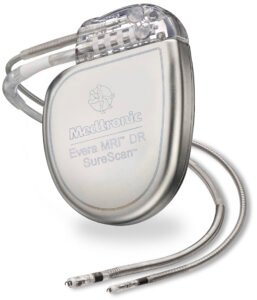
Table of contents
- 1. Why the heart needs an ICD
- 2. Working principle: the ICD as a control loop
- 3. Device architecture and components
- 4. Subcutaneous vs. transvenous vs. extravascular: comparison
- 5. Detection algorithms
- 6. Therapy delivery
- 7. Implantation procedure
- 8. Biomaterials and power source
- 9. Complications and failure modes, with real numbers
- 10. QA, testing, and follow-up parameters
- 11. Regulatory and standards framework
- 12. Manufacturer landscape (representative, non-exhaustive)
- 13. Future directions
- 14. Conclusion
- FAQ
- References
A practical, numbers-first guide for biomedical engineers — physiology, architecture, detection logic, implantation, QA/testing, complications, and where the technology is heading, built on verified 2025–2026 literature.
1. Why the heart needs an ICD
Sudden cardiac death is most often caused by ventricular fibrillation (VF) — a chaotic, disorganized electrical activity in the ventricles that replaces an effective pump with a quiver, and by sustained ventricular tachycardia (VT) — a fast, often regular rhythm originating from abnormal ventricular tissue (commonly a scar from a prior heart attack) that can degenerate into VF. Both stop effective blood flow to the brain and body within seconds to a couple of minutes. External defibrillation — the paddles/pads used by paramedics or an AED — works, but only if a rhythm check happens and a shock is delivered within that narrow window, which is rarely fast enough for an event that occurs at home or during sleep.
An ICD solves the timing problem by keeping the “electrode and decision-maker” permanently attached to the heart. It can detect a dangerous rhythm and deliver therapy within seconds of onset, without waiting for anyone to notice.
2. Working principle: the ICD as a control loop
At its core, an ICD runs the same three-stage loop, continuously, for years:
- Sense — one or more electrodes on the lead(s) pick up the intracardiac electrogram (a much cleaner, higher-amplitude signal than a surface ECG because the electrode sits directly on or in the heart muscle).
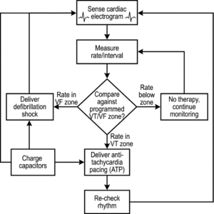
- Classify — onboard firmware measures the interval between consecutive sensed beats (rate) and, in most modern devices, also analyzes the shape of the electrical signal (morphology) and its regularity/onset pattern, then compares these against programmed zones (e.g., a “VT zone” and a faster “VF zone”) to decide whether the rhythm is a treatable tachyarrhythmia or a benign fast rhythm (like sinus tachycardia during exercise) that should not be shocked.
- Act — if therapy is warranted, the device chooses between anti-tachycardia pacing (ATP) for a hemodynamically tolerated VT, or a full defibrillation shock for VF or a VT that doesn’t respond to ATP, and delivers it through the same or a dedicated shocking electrode.
The critical engineering tension is between sensitivity (never missing a true VF, since a missed episode can be fatal) and specificity (never shocking a benign rhythm, since inappropriate shocks are painful, psychologically traumatic, and can themselves provoke arrhythmia). Across the ICD population, roughly 10–15% of patients experience at least one inappropriate shock over their device’s lifetime, most commonly from rhythm misclassification (e.g., supraventricular tachycardia mistaken for VT) or lead-related oversensing4 — which is why so much of the ICD’s internal logic exists purely to raise specificity without sacrificing the sensitivity that makes the device worth implanting in the first place.
3. Device architecture and components
An ICD system has two physical parts: the pulse generator (the “can”) and one or more leads.
3.1 The pulse generator
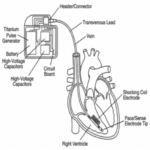
- Hermetically sealed titanium can housing the battery, high-voltage capacitors, and the circuit board (microcontroller, sensing amplifiers, high-voltage charging/output circuitry, and telemetry radio).
- Battery: primary (non-rechargeable) lithium chemistry — most commonly lithium/silver vanadium oxide (Li/SVO) or a lithium/manganese-dioxide hybrid — chosen because it can deliver both a small continuous current for sensing/pacing and, on demand, a large pulse of current to charge the high-voltage capacitors in a few seconds. Typical device longevity is around 5–7 years for a single-chamber or dual-chamber ICD, shorter for devices that pace frequently or deliver many shocks.1
- High-voltage capacitors: store the charge for the defibrillation shock; charging them from the battery before a shock takes a few seconds, which is why an ICD “delays” a fraction before a defibrillation shock but not before a pacing pulse.
- Telemetry: short-range wireless link used by the clinic programmer and, in most current devices, a home remote-monitoring transmitter that sends alerts (e.g., battery status, detected arrhythmia episodes, lead impedance trends) to the clinical team between visits.
3.2 Leads and lead architecture
Leads are insulated, multi-conductor wires that carry sensing and pacing/shock current between the can and the heart. Depending on the architecture, an ICD may use:
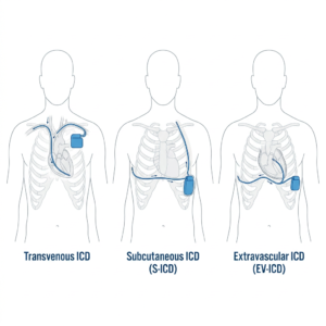
- Transvenous ICD (TV-ICD): one or more leads threaded through the subclavian (or other central) vein into the right ventricle (single-chamber) and optionally the right atrium (dual-chamber) or a coronary-sinus branch for the left ventricle (cardiac resynchronization-defibrillator, CRT-D). The right-ventricular lead carries a shocking coil in addition to standard pace/sense electrodes, and fixation is either passive (tines that catch in the trabeculae) or active (a screw-in helix), each with different perforation-risk profiles discussed in Section 9.
- Subcutaneous ICD (S-ICD): no lead touches the heart or the venous system at all. A single lead runs subcutaneously along the sternum, and the pulse generator sits laterally under the arm; sensing and shocking both happen from outside the rib cage.5
- Extravascular ICD (EV-ICD): an emerging third architecture (FDA-cleared devices reaching wider clinical use in 2024–2026) that tunnels a lead into the substernal space — outside the heart and blood vessels but still within the chest — aiming to combine the shock efficacy and pacing capability of a transvenous system with the lower vascular-complication profile of a subcutaneous one.27
4. Subcutaneous vs. transvenous vs. extravascular: comparison
| Feature | Transvenous (TV-ICD) | Subcutaneous (S-ICD) | Extravascular (EV-ICD) |
|---|---|---|---|
| Lead location | Inside heart chambers, via veins | Under the skin, outside ribs/heart | Substernal, outside vessels/heart |
| Vascular/intracardiac complications | Present (thrombosis, perforation, endocarditis) | Eliminated | Eliminated |
| Anti-tachycardia pacing (ATP) | Yes, native | Historically no; newer systems add ATP via a paired leadless pacemaker using unidirectional conductive communication36 | Yes, native |
| Bradycardia (backup) pacing | Yes | No (pause-only shock therapy) | Limited |
| Defibrillation energy | Typically ~30–40 J | Higher output needed (larger inter-electrode distance) | Comparable to TV-ICD |
| Best fit | Patients needing pacing support or CRT | Patients with vascular access issues, prosthetic valves, or younger patients wanting to avoid intravascular hardware | Emerging option positioned between the two |
The PRAETORIAN trial (2025 follow-up analysis) found the S-ICD non-inferior to TV-ICDs for device-related complications and inappropriate-shock rates, reinforcing it as a mainstream option rather than a niche one.5
5. Detection algorithms
Detection is the single most engineered part of an ICD, because a wrong call in either direction has serious consequences. Modern algorithms combine several discriminators, run in this general sequence:
- Rate/interval measurement against programmed zone boundaries (e.g., “VT zone” starting around 150–180 bpm, “VF zone” above that, both individually programmed per patient by the treating physician).
- Onset pattern — a sudden rate jump is more suspicious for a pathological tachyarrhythmia than a gradual acceleration typical of sinus tachycardia during exertion.
- Stability — beat-to-beat interval variability; atrial fibrillation with rapid, irregular conduction to the ventricles looks different from a regular monomorphic VT.
- Morphology/electrogram-shape comparison against a stored template of the patient’s normal QRS-like signal — a big deviation suggests the beat originates from an abnormal ventricular focus rather than the normal conduction pathway.
- Dual-chamber discrimination (when an atrial lead is present) — comparing atrial and ventricular rates/timing helps separate supraventricular tachycardias from true VT.
These layered discriminators (proprietary combinations sold under names like PR Logic™ and similar manufacturer-specific algorithm families) are the direct descendants of decades of inappropriate-shock data, and 2023–2025 literature is now actively exploring machine-learning-based rhythm classifiers to push specificity further, though these require extensive clinical validation before being embedded in a life-critical device.4
6. Therapy delivery
Once a treatable rhythm is confirmed, the ICD chooses among:
- Anti-tachycardia pacing (ATP): a short burst of low-energy pacing pulses (roughly 0.5–4 V amplitude) delivered faster than the tachycardia’s own rate, aiming to capture and interrupt the reentrant electrical circuit that sustains the VT — painless when it works, and the preferred first-line therapy for hemodynamically tolerated VT because it avoids a shock altogether.3
- Cardioversion: a synchronized, lower-energy shock timed to a sensed beat, used for some organized VTs that don’t respond to ATP.
- Defibrillation: a high-energy, unsynchronized shock — typically in the range of about 30–40 joules delivered at maximum output — used for VF or a VT that has degenerated or failed ATP/cardioversion. This is the shock patients describe as feeling like “being kicked in the chest,” and it is the therapy that actually terminates VF, restoring an organized rhythm the heart’s own pacemaker cells can then take back over.1
7. Implantation procedure
TV-ICD implantation is typically performed under local anesthesia with conscious sedation (rarely general anesthesia), in a cardiac catheterization or electrophysiology lab, and follows roughly this sequence:
- Venous access (usually subclavian or axillary vein) and lead advancement under fluoroscopic guidance into the right ventricle (and right atrium/coronary sinus branch if dual-chamber or CRT-D).
- Lead fixation (passive tines or active screw-in helix) and measurement of sensing amplitude, pacing threshold, and lead impedance to confirm good electrical contact.
- Formation of a subcutaneous or sub-pectoral pocket for the pulse generator, typically below the collarbone.
- Connection of the lead(s) to the generator header and defibrillation threshold testing (inducing a brief VF episode and confirming the device detects and terminates it) — increasingly omitted in routine cases given modern devices’ reliable safety margins, but still used selectively.
- Wound closure, followed by a chest X-ray to confirm lead position and rule out pneumothorax, and device interrogation to store final baseline parameters.
S-ICD implantation differs substantially: no fluoroscopy or venous access is required; the generator is placed laterally, and the lead is tunneled subcutaneously along the sternum, making the whole procedure attractive for patients where venous access is difficult or undesirable.
8. Biomaterials and power source
- Can: titanium, chosen for biocompatibility, mechanical strength at thin wall sections, and hermeticity.
- Header/connector block: medical-grade epoxy or polyurethane, molded to accept the international standard (DF-1/DF-4 or IS-1) lead connectors.
- Lead insulation: silicone rubber or polyurethane, chosen for flexibility and long-term fatigue resistance under the roughly 100,000+ flex cycles a lead experiences per day from the beating heart.
- Electrode tips: platinum-iridium alloy, valued for corrosion resistance and stable, low-polarization sensing/pacing performance over years.
- Battery: lithium/silver vanadium oxide (Li/SVO) remains the dominant chemistry specifically because it supports both the low continuous drain of sensing/pacing and the high pulsed current needed to charge the shock capacitors — a combination most other battery chemistries can’t deliver in one cell.1
9. Complications and failure modes, with real numbers
Acute/procedural:
– Lead perforation: roughly 0.1–0.8% incidence, most often at the right-ventricular free wall; septal or left-ventricular perforation is rarer but can be life-threatening, presenting as cardiac tamponade, pericardial effusion, or hemothorax. Risk rises with active-fixation leads, over-torquing during implantation, right-ventricular septal lead positioning, and in patients who are female, older, have thin ventricular walls, COPD, or a low BMI.4
– Pneumothorax and vascular access injury during venous puncture.
Lead-related (transvenous only):
– Device/lead infection or endocarditis: roughly 0.5–2% annually.
– Lead dislodgment, insulation failure, and conductor fracture — the classic long-term transvenous-lead failure modes that the S-ICD and EV-ICD architectures were specifically designed to avoid.
– Venous thrombosis around the lead.
Device-related, architecture-independent:
– Battery depletion at end of service life, requiring generator replacement.
– Inappropriate shocks from oversensing (e.g., electromagnetic interference, lead fracture noise, or T-wave oversensing) or rhythm misclassification — the 10–15% inappropriate-shock rate referenced in Section 2, with meaningful associated psychological distress and quality-of-life impact.4
– Lead perforation can, less commonly, present weeks to years after implantation due to gradual myocardial remodeling around the fixation point, not only acutely.4
Device recalls remain a real operational risk in this category: 2024 recalls affecting Medtronic’s Cobalt line and Boston Scientific’s EMBLEM S-ICD together affected more than 100,000 implanted devices, underscoring why post-market surveillance and remote monitoring matter as much as initial implant quality.12
10. QA, testing, and follow-up parameters
Manufacturing/acceptance-level QA (representative categories, verified against each device’s own IFU rather than assumed):
– Hermeticity (helium leak testing of the can).
– Battery capacity and internal-impedance verification.
– High-voltage capacitor charge-time and output-energy accuracy across the rated range.
– Lead pull-strength, insulation-integrity, and flex-fatigue testing to the relevant cycle counts.
– Electromagnetic compatibility testing against the device’s labeled MRI-conditional status, when applicable.
Follow-up (in-service) checks at each clinic visit or remote transmission, compared against the previous baseline rather than an absolute threshold:
– Battery voltage/remaining longevity estimate.
– Lead impedance (a sudden jump or drop can flag a fracture or insulation breach).
– Sensing amplitude and pacing threshold (rising thresholds can indicate lead maturation, fibrosis, or a developing problem).
– Stored arrhythmia episodes and delivered therapies (ATP vs. shock counts), reviewed for appropriateness.
– Remote-monitoring alert history between in-person visits — now standard practice, and one of the main levers for catching a developing lead or battery problem before it becomes symptomatic.
11. Regulatory and standards framework
- FDA (United States): ICDs are Class III devices requiring premarket approval (PMA), with mandatory postmarket adverse-event reporting (MAUDE database) and, when needed, recall authority — the mechanism behind the 2024 Cobalt/EMBLEM recalls referenced above.12
- IEC 60601 series: general electrical safety and essential-performance standards for medical electrical equipment, with device-specific defibrillator provisions (e.g., within the IEC 60601-2-4 family) covering output accuracy and safety.
- ISO 14708 series: implants for surgery — active implantable medical devices — the baseline safety/performance standard family for devices like ICDs and pacemakers.
- EU MDR (2017/745): ICDs fall under the highest-risk implantable-device classification in the EU, requiring notified-body conformity assessment and post-market clinical follow-up.
- AHA/ACC guidelines: clinical (not device-safety) guidance defining which patients meet indications for ICD implantation, used by clinicians and payers (including CMS coverage policy) alongside the regulatory framework.8
12. Manufacturer landscape (representative, non-exhaustive)
| Manufacturer | Headquarters | Notable position (2024–2025) |
|---|---|---|
| Medtronic | Dublin, Ireland | Broad TV-ICD/CRT-D portfolio; Aurora EV-ICD received FDA clearance in 2024, expanding into the extravascular category12 |
| Boston Scientific | Marlborough, MA, USA | Market-share leader in the combined US cardiac rhythm management/EP/ablation market; its EMBLEM S-ICD system received favorable US coding/payment designations in 2024910 |
| Abbott | Chicago, IL, USA | Major TV-ICD and CRT-D portfolio (legacy St. Jude Medical lines) |
| Biotronik | Berlin, Germany | Active across transvenous and subcutaneous ICD segments |
| LivaNova | London, UK | Smaller cardiac rhythm management portfolio presence11 |
Exact current battery-longevity, output-energy, and dimension specifications vary by model and are published in each manufacturer’s technical manual/IFU; this guide intentionally reports only the ranges independently confirmed across the cited literature rather than a single vendor’s marketing figures.
13. Future directions
- Extravascular ICD (EV-ICD) maturation: 2025–2026 clinical data continues to show high defibrillation success and effective ATP delivery with a lower complication profile than transvenous systems, positioning EV-ICD as a serious middle path between TV-ICD and S-ICD.27
- S-ICD/leadless-pacemaker pairing: newer systems add ATP capability to subcutaneous defibrillation by having the S-ICD wirelessly command a separately implanted leadless pacemaker via unidirectional conductive communication — closing the historical functional gap between S-ICD and TV-ICD.36
- AI-assisted rhythm discrimination: ongoing work on machine-learning-based classifiers aimed at reducing the inappropriate-shock rate without sacrificing sensitivity to true VF/VT, still in clinical-validation stages as of 2023–2025 literature.4
- Expanded remote monitoring: deeper integration of continuous home telemetry for earlier detection of lead or battery issues between scheduled visits.
14. Conclusion
An ICD is, functionally, a permanently implanted rhythm-classification computer wired directly to the heart, built to make a life-or-death decision — pace quietly, shock hard, or do nothing — in under a few seconds, correctly, for years on a single battery. Its engineering story is really the story of narrowing the gap between sensitivity (never missing true VF) and specificity (never shocking a benign rhythm), across three now-competing lead architectures (transvenous, subcutaneous, extravascular) that trade off pacing capability against vascular risk in different ways.
FAQ
Is an ICD the same as a pacemaker?
No. A pacemaker mainly treats a heart rate that is too slow. An ICD’s primary role is detecting and stopping a dangerously fast, potentially fatal rhythm (VT/VF), though many ICDs also include backup pacemaker functionality for slow rhythms.
Does getting shocked by an ICD hurt?
Yes — a defibrillation shock is often described as a sudden, brief, forceful jolt, sometimes compared to being kicked in the chest. Anti-tachycardia pacing, used first when appropriate, is painless.
How long does an ICD battery last?
Typically around 5–7 years, depending on how much pacing and how many shocks the device delivers; the device itself tracks remaining battery status and alerts the care team before replacement is needed.1
Can everyone get a subcutaneous ICD instead of a transvenous one?
Not everyone — historically S-ICDs couldn’t deliver anti-tachycardia pacing or backup bradycardia pacing, though newer paired systems are narrowing that gap. The right choice depends on a patient’s specific rhythm history, vascular anatomy, and pacing needs, decided by the treating electrophysiologist.56
References
- Mahmoodi M, et al. (2026). Cardiac defibrillators in modern healthcare: a review of functionality, safety, and future directions. Bulletin of the National Research Centre. DOI: 10.1186/s42269-026-01420-z ↩↩↩↩
- Collings S, et al. (2026). Preservation of an extravascular implantable cardioverter-defibrillator. Journal of Cardiovascular Electrophysiology / ScienceDirect. https://www.sciencedirect.com/science/article/pii/S2214027126000825 ↩↩
- Pujol-Lopez M, Tung R, Mont L. (2026). Innovations in cardiac device therapy in the era of advanced rhythm management: implantable defibrillators and conduction system pacing. Heart. DOI: 10.1136/heartjnl-2025-325834 ↩↩↩
- Alexiou P, Ballas CE, Alexiou C, et al. (2026). Implantable Cardioverter-Defibrillator (ICD) Lead-Induced Septal and Left Ventricular Perforation in Hypertrophic Cardiomyopathy: A Case Report. Cureus, 18(2):e103333. DOI: 10.7759/cureus.103333 (PMC12981612) ↩↩↩↩↩↩
- Hauser RG, Desouki M, Stanberry LI, et al. (2026). Inappropriate shocks from subcutaneous and transvenous implantable cardioverter-defibrillators: Reports from the FDA MAUDE database. Heart Rhythm O2. DOI: 10.1016/j.hroo.2025.11.002 (PMC12925796) ↩↩↩
- Angelini E, Albert K, Duncker D, et al. (2025). The extravascular implantable cardioverter-defibrillator: technology, evidence, and clinical perspectives. Herz, 50(6):417-426. (PMC12660413) ↩↩↩
- Markman TM, et al. (2026). The extravascular ICD: Evolving paradigms in sudden cardiac death prevention. Heart Rhythm Journal. https://www.heartrhythmjournal.com/article/S1547-5271(25)02931-5/fulltext ↩↩
- FEP Blue. (2025). Medical Policy 7.01.44 — Implantable Cardioverter Defibrillators. PDF ↩
- Mordor Intelligence. (2025). Defibrillator Companies — Key Players & Market Share. https://www.mordorintelligence.com/industry-reports/defibrillator-market/companies ↩
- iData Research. (2025). United States Cardiac Rhythm Management Market Report. https://idataresearch.com/product/cardiac-rhythm-management-market-united-states/ ↩
- Market Research Future. (2024–2025). Pacemaker Companies and CRM Market Leaders. https://www.marketresearchfuture.com/reports/pacemaker-market/companies ↩
- Mordor Intelligence. (2024). Implantable Defibrillators Market — Recalls and FDA Clearances. https://www.mordorintelligence.com/industry-reports/implantable-defibrillators-market ↩↩↩