How Does a Pulse Oximeter Work? Photoplethysmography and SpO2 Explained
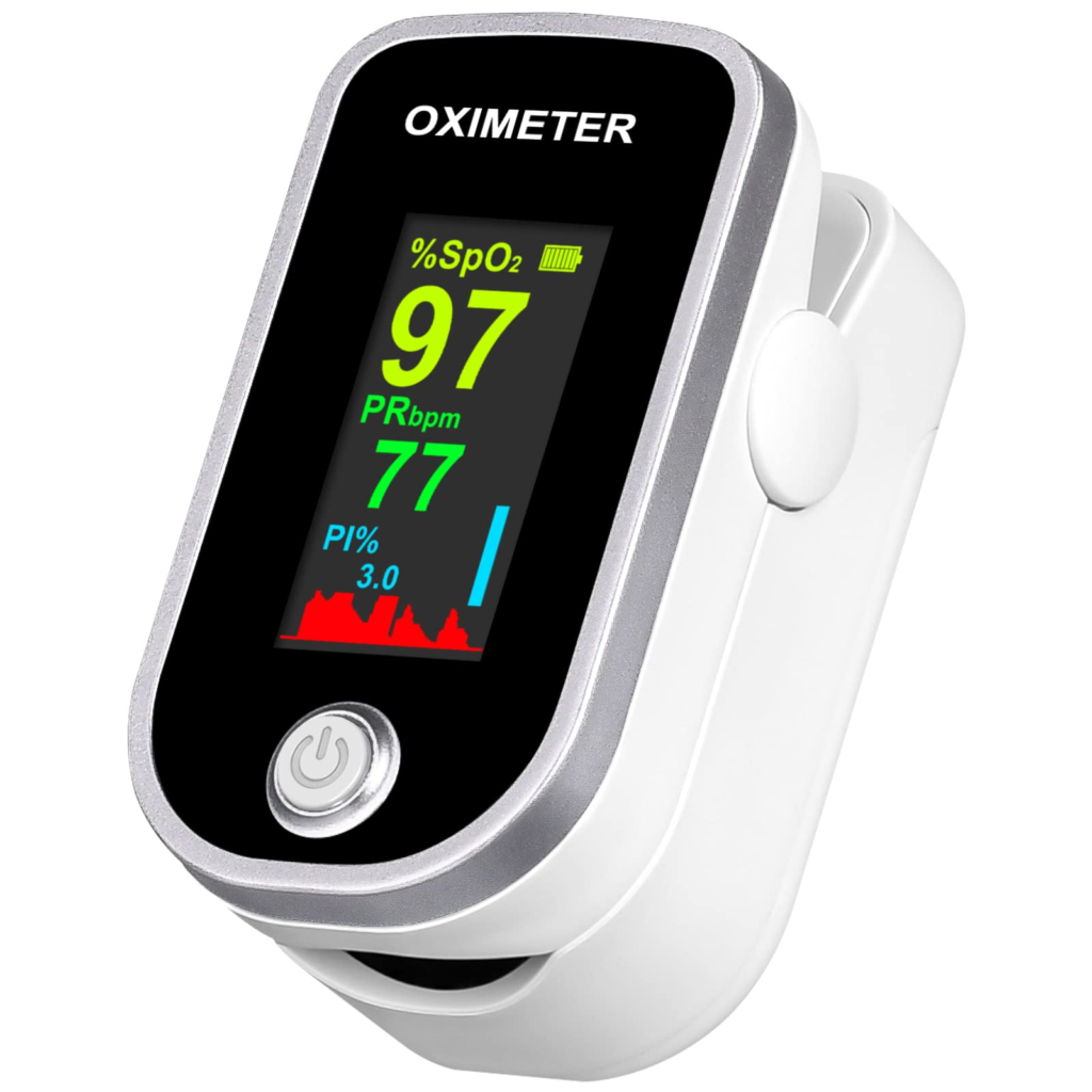
A pulse oximeter is a small, non-invasive device — usually a clip worn on a fingertip, earlobe, or forehead — that estimates the percentage of oxygen-carrying hemoglobin in arterial blood, a value called SpO2 (peripheral oxygen saturation), along with pulse rate, without ever drawing blood. It works by shining two wavelengths of light — red and infrared — through (or off) living tissue and measuring how much of each wavelength is absorbed. Oxygenated and deoxygenated hemoglobin absorb these two wavelengths differently, and the tiny, rhythmic changes in absorption caused by each heartbeat’s pulse of arterial blood let the device isolate the arterial signal from the surrounding skin, bone, and venous blood and calculate an oxygen saturation estimate — a technique built on optical physics called photoplethysmography (PPG) combined with the Beer-Lambert law of light absorption.
Everything below unpacks that definition — starting from why continuous, non-invasive oxygen monitoring is needed, through the optical and signal-processing engineering behind the SpO2 calculation, how the device is built, operated, tested, maintained, and where the field is heading next.
1. Why pulse oximetry is needed
Adequate blood oxygen levels are essential for every organ in the body, and a drop in oxygen saturation (hypoxemia) can happen silently, well before a patient looks or feels distressed. Before pulse oximetry became widespread, clinicians relied mainly on arterial blood gas sampling — an invasive, single-point-in-time test — to know a patient’s oxygenation status. A continuous, non-invasive, essentially instant readout of SpO2 and pulse rate transformed anesthesia monitoring, emergency and critical care, neonatal care, sleep studies, and home monitoring for chronic respiratory disease, giving clinicians (and increasingly patients themselves) an early-warning signal for deteriorating oxygenation.
2. Working principle: from two LEDs to an SpO2 number
2.1 Photoplethysmography — sensing the arterial pulse optically
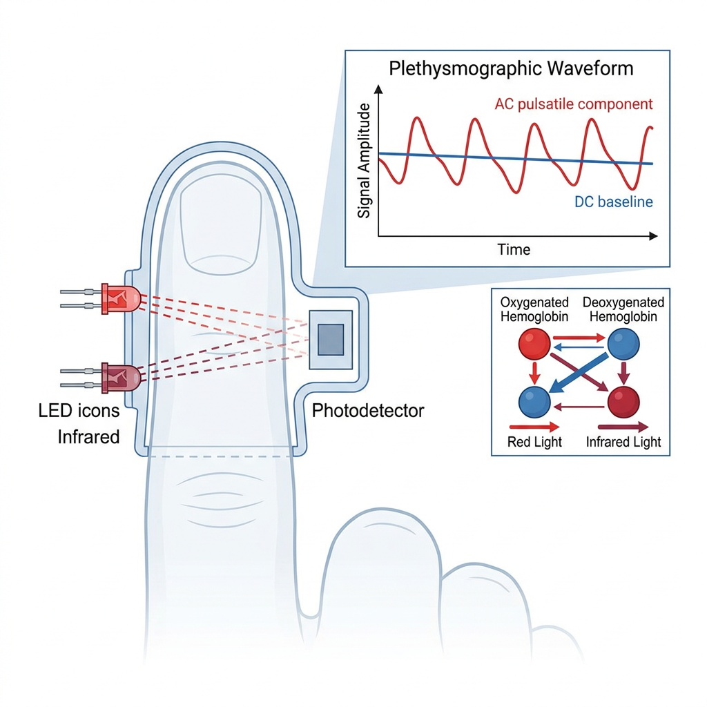
Photoplethysmography (PPG) is the underlying optical technique: light is shone into tissue, and a photodetector on the other side (transmission mode, the classic finger-clip design) or on the same side (reflectance mode, common in wearables) measures how much light gets through or bounces back. That measured light intensity has two components — a DC component, the large, slowly changing baseline absorption from skin, bone, tissue, and the non-pulsating (venous and capillary) blood volume, and a much smaller AC component, the rhythmic ripple in absorption caused by the pulsing arterial blood volume that expands and contracts with every heartbeat. Isolating that small AC ripple from the much larger DC baseline is what lets the device measure a signal that specifically reflects arterial blood — the same blood a blood gas sample would measure — rather than an average across all the tissue types the light passes through.
2.2 The Beer-Lambert law and the “ratio of ratios”
The amount of light absorbed as it passes through a substance follows the Beer-Lambert law:
A = log(I₀ / I) = E × C × D
where A is absorbance, I₀ is the incident light intensity, I is the transmitted light intensity, E is the wavelength-specific extinction coefficient of the absorbing substance (here, hemoglobin), C is the concentration of that substance, and D is the thickness of tissue the light travels through.
Oxygenated hemoglobin (O₂Hb) and deoxygenated hemoglobin (RHb) have different extinction coefficients at red (~660 nm) and infrared (~930–940 nm) wavelengths: red light is absorbed considerably more by deoxygenated hemoglobin, while infrared light is absorbed nearly equally by both — which is why a well-oxygenated finger reading looks different in transmitted red light than in infrared. By measuring the change in absorbance (ΔA) at both wavelengths across just the pulsatile (AC) component, the device computes a “ratio of ratios” — the ratio of the red AC/DC absorbance ratio to the infrared AC/DC absorbance ratio — which is mathematically related to the fraction of hemoglobin that is oxygenated. Because that ratio cancels out most of the DC baseline (tissue thickness, skin pigment, and other non-pulsatile absorbers), the calculation is far more robust to individual anatomical differences than a simple raw-intensity measurement would be. The device converts the ratio-of-ratios value to an SpO2 percentage using an empirical calibration curve, built from reference measurements against arterial blood gas co-oximetry during clinical validation studies rather than a purely theoretical formula.
2.3 Two sensor geometries
- Transmission mode — the light source and photodetector are on opposite sides of the tissue (the classic spring-loaded fingertip clip); light passes fully through the tissue bed. This is the standard clinical configuration because the transmitted signal is generally stronger and less affected by ambient light.
- Reflectance mode — the light source and photodetector sit side by side on the same surface, measuring light scattered back from the tissue; used in forehead sensors and most wrist-worn or other wearable devices where a full transmission path isn’t practical.
3. Device architecture and components
- Dual-wavelength LED emitter — typically a red LED (~660 nm) and an infrared LED (~930–940 nm), rapidly alternated (multiplexed) so the photodetector can attribute each light pulse to its source wavelength; a third wavelength is sometimes added on higher-end platforms for improved accuracy or additional parameters.
- Photodetector (photodiode) — converts the transmitted or reflected light into an electrical signal proportional to received light intensity.
- Analog front-end and amplification — amplifies and filters the small photodetector signal before digitization; ambient light rejection and gain control compensate for varying tissue thickness and skin tone across patients.
- Signal processing unit / microcontroller — digitizes the amplified signal and runs the algorithms that separate AC from DC components, compute the ratio of ratios, apply the calibration curve to output SpO2, calculate pulse rate from the pulsatile waveform period, and run motion-artifact reduction algorithms.
- Motion-artifact reduction algorithms — adaptive digital filters, such as normalized least mean squares (NLMS) or recursive least squares (RLS) algorithms, that use the pulsatile waveform’s known characteristics (and, on many modern platforms, an onboard accelerometer) to distinguish a genuine pulse from motion-induced noise; a high-pass filter around 0.1 Hz removes the DC baseline drift and a low-pass filter around 30 Hz removes high-frequency electrical noise.
- Display and alarm system — shows numeric SpO2 and pulse rate, the plethysmographic (pulse) waveform, and triggers audible/visual alarms when readings breach clinician-set thresholds; hospital-grade monitors and standalone fingertip devices vary widely in display and alarm sophistication.
- Sensor/probe and cable — the reusable or single-patient-use clip, adhesive wrap, or wearable housing containing the LEDs and photodetector, connected to the monitor by a cable (or wirelessly, on some wearable platforms).
4. Classification
| Category | Type | Key characteristic |
|---|---|---|
| Sensor geometry | Transmission-mode | Classic fingertip/earlobe clip; light passes through tissue |
| Sensor geometry | Reflectance-mode | Forehead or wrist-worn sensor; light reflects back from tissue |
| Use setting | Standalone fingertip device | Battery-powered, self-contained display, spot-check or short-term home use |
| Use setting | Bedside/patient monitor module | Continuous monitoring, integrated alarms, often multi-parameter (with ECG, capnography, etc.) |
| Use setting | Wearable/consumer PPG device | Continuous ambulatory monitoring, often paired with a smartphone app; accuracy and regulatory status vary widely by product |
5. Step-by-step: taking a pulse oximetry reading
- Select and prepare the site. A finger, toe, or earlobe (or forehead for reflectance sensors) is chosen with adequate perfusion; nail polish, artificial nails, and cold or poorly perfused extremities are avoided or noted, as they can distort readings.
- Apply the sensor. The clip or wrap is applied snugly but not so tight as to restrict blood flow, aligning the LED emitter and photodetector directly across from (transmission) or beside (reflectance) each other.
- Allow signal stabilization. The device typically takes several seconds to a small number of pulse cycles to acquire a stable plethysmographic waveform before displaying a reliable reading.
- Read SpO2, pulse rate, and waveform quality. The clinician checks the numeric SpO2 and pulse rate against the patient’s clinical status and confirms the displayed pulse waveform looks physiologic (not flat or erratic), since a poor waveform undermines confidence in the numeric reading.
- Reposition or investigate if signal quality is poor. Motion, poor perfusion, ambient light, or sensor misplacement are corrected before trusting a low or erratic reading — a suspiciously low SpO2 in an otherwise well patient warrants rechecking the sensor before treating it as a true clinical finding.
- Set alarm limits (continuous monitoring). For continuous bedside use, clinician-appropriate high/low SpO2 and pulse-rate alarm thresholds are configured according to the patient’s clinical situation.
6. Sensing, control, and stimulation parameters
| Parameter | Typical value/range | Notes |
|---|---|---|
| Red wavelength | ~660 nm | Absorbed considerably more by deoxygenated hemoglobin |
| Infrared wavelength | ~930–940 nm | Absorbed nearly equally by oxy- and deoxyhemoglobin |
| SpO2 measurement range | Typically validated 70–100% | Standard’s accuracy claims are validated across this range |
| Accuracy metric | ARMS (Accuracy Root Mean Square) | Current international standard ceiling commonly ~3.5–4% ARMS; regulators have proposed tightening this limit |
| High-pass filter cutoff | ~0.1 Hz | Removes DC baseline drift |
| Low-pass filter cutoff | ~30 Hz | Removes high-frequency electrical noise |
7. QA and safety-testing requirements
- Governing standard: ISO 80601-2-61 — the international particular standard for the basic safety and essential performance of pulse oximeter equipment, specifying required accuracy validation, alarm behavior, essential performance during motion, and signal dropout/recovery testing.
- Accuracy validation (ARMS) — devices must demonstrate their measured SpO2 accuracy against reference co-oximetry measurements as an Accuracy Root Mean Square (ARMS) value; current typical accepted limits are in the ~3.5–4% ARMS range, and regulators (including the FDA, per public docket FDA-2023-N-4976) have proposed tightening these limits further, along with requiring more diverse skin-pigmentation representation in validation studies.
- Performance under motion and low perfusion — the standard requires testing device behavior during motion artifact and low-perfusion conditions, since these are the settings where pulse oximeter readings are historically least reliable.
- General electrical safety — pulse oximeters, like other medical electrical equipment, are also tested against IEC 60601-1 general basic safety and essential performance requirements.
- Skin tone / pigmentation performance — growing clinical and regulatory attention has focused on documented reduced accuracy of pulse oximetry in patients with darker skin pigmentation, driving updated validation requirements calling for more diverse clinical study populations.
8. Maintenance and troubleshooting
| Interval | Task | Acceptance criteria |
|---|---|---|
| Before each use | Inspect sensor/probe for cracks, discoloration, or damaged cable | Clean optical windows, no visible LED/photodetector damage, secure cable connection |
| Before each use | Confirm correct sensor size/type for the patient (adult/pediatric/neonatal) | Sensor fits the intended site without excessive pressure or gapping |
| Periodically | Clean reusable sensors per manufacturer instructions | No residue over the optical windows that could scatter or block light |
| Periodically | Battery check (standalone devices) | Sufficient charge for the intended monitoring duration |
| Per manufacturer schedule | Biomedical/clinical engineering functional check against a calibrated simulator | Displayed SpO2 and pulse rate match the simulator’s reference output within specification |
| Failure mode | Typical signature | Usual cause |
|---|---|---|
| No reading / “searching” indefinitely | Blank or dashed SpO2 display | Poor sensor placement, very low perfusion, or ambient light interference |
| Erratic or rapidly fluctuating readings | Numeric value jumps or waveform looks noisy | Patient motion, poor sensor fit, or shivering |
| Falsely low SpO2 in a clinically well patient | Low numeric reading with a good-looking waveform | Nail polish/artificial nails, cold extremity, sensor misplacement, or (documented in the literature) reduced accuracy related to skin pigmentation |
| Falsely normal SpO2 in a genuinely hypoxic patient | Normal-looking reading despite clinical deterioration | Carbon monoxide poisoning (standard 2-wavelength devices cannot distinguish carboxyhemoglobin from oxyhemoglobin) or methemoglobinemia |
9. Complications, safety, and limitations
- Cannot detect carbon monoxide poisoning reliably. Standard two-wavelength pulse oximeters cannot distinguish carboxyhemoglobin from oxyhemoglobin and can display a falsely reassuring normal SpO2 in a patient with significant carbon monoxide poisoning; specialized multi-wavelength co-oximeters are needed for that distinction.
- Skin pigmentation-related accuracy differences. Multiple clinical studies have documented reduced accuracy — particularly a tendency toward falsely normal readings in patients with clinically significant hypoxemia — in patients with darker skin pigmentation, an active area of regulatory and manufacturer attention.
- Motion and low-perfusion artifact. Both remain the most common practical source of unreliable readings, even with modern adaptive-filtering algorithms.
- Nail polish, artificial nails, and interfering substances. Certain nail polish colors (especially dark blue, green, and black) and some artificial nails can absorb or scatter light abnormally and distort readings.
- Not a substitute for full oxygenation/ventilation assessment. SpO2 reflects oxygen saturation, not ventilation (CO2 clearance) — a patient can have a normal SpO2 while under-ventilating, particularly when supplemental oxygen is being given.
- Pressure injury risk. Prolonged use of a clip-style sensor on the same digit can cause localized pressure injury; site rotation is recommended for continuous long-term monitoring.
10. Regulatory and standards framework
- ISO 80601-2-61 — international particular standard for the basic safety and essential performance of pulse oximeter equipment.
- IEC 60601-1 — the general basic safety and essential performance standard for medical electrical equipment that ISO 80601-2-61 supplements.
- US regulatory pathway — pulse oximeters intended for medical use are regulated by the FDA, generally as Class II devices requiring 510(k) clearance; the FDA has an active public docket (FDA-2023-N-4976) addressing proposed updates to accuracy and skin-pigmentation-related performance requirements.
- Consumer/wellness devices — many wrist-worn or app-based PPG products are explicitly marketed as “wellness” rather than medical devices, exempting them from the same clinical accuracy validation that a medical-grade pulse oximeter must pass — a distinction clinicians and buyers should not overlook.
11. Manufacturer landscape (representative, non-exhaustive)
| Manufacturer | Representative technology | Notable specifications |
|---|---|---|
| Masimo | Signal Extraction Technology (SET) | In-motion accuracy reported at approximately 1.5% ARMS in motion, using multiple wavelengths and adaptive noise cancellation techniques aimed specifically at low-perfusion and motion conditions |
| Medtronic (Nellcor OxiMax) | OxiMax algorithm platform | In-motion accuracy reported at approximately 3% ARMS in motion; long clinical track record, with published comparative accuracy studies against other platforms in critically ill patients |
| Nonin | Standalone/portable pulse oximetry | Widely used in portable fingertip and handheld clinical devices; exact ARMS specifications vary by model and should be confirmed against the current product datasheet |
Exact accuracy specifications, validated SpO2 range, and motion-tolerance claims vary by specific model and firmware/sensor combination — always verify the current published datasheet before a procurement or clinical decision.
12. Comparison: pulse oximetry vs. related oxygenation/ventilation monitoring
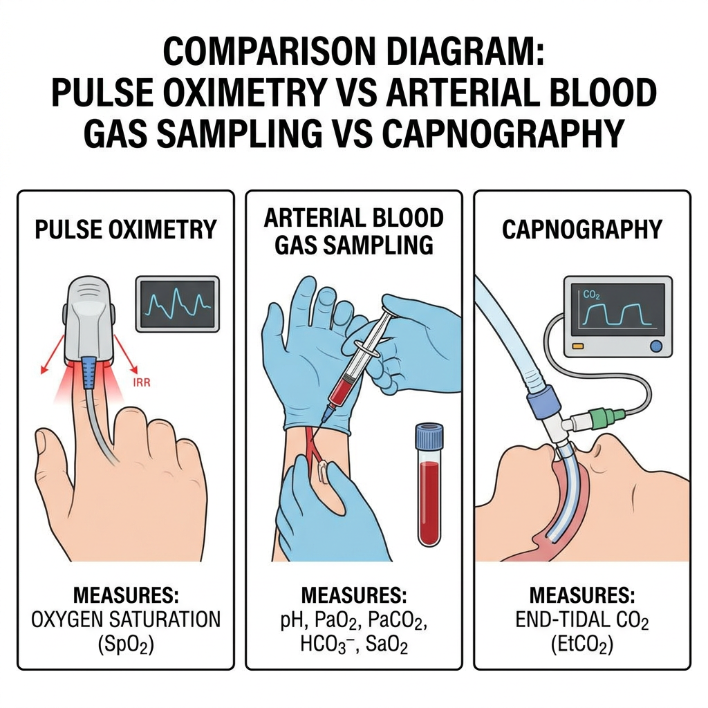
| Factor | Pulse oximetry (SpO2) | Arterial blood gas (ABG) sampling | Capnography (EtCO2) |
|---|---|---|---|
| What it measures | Percentage of hemoglobin saturated with oxygen, estimated optically | Directly measured PaO2, PaCO2, pH, and other blood gas values from a drawn sample | End-tidal carbon dioxide, reflecting ventilation adequacy |
| Invasiveness | Non-invasive, continuous | Invasive, single point in time (arterial puncture) | Non-invasive, continuous (via airway sampling) |
| What it does NOT tell you | Ventilation adequacy (CO2 clearance); cannot reliably flag carbon monoxide poisoning | Requires clinical staff and lab turnaround; not continuous | Oxygenation status directly |
| Typical use case | Continuous bedside/ambulatory oxygenation monitoring | Definitive, precise blood gas values when SpO2 alone is insufficient | Continuous ventilation monitoring, especially under sedation/anesthesia |
13. Future directions
Tighter, more diverse accuracy standards. Regulatory efforts already underway (e.g., the FDA’s public docket on ARMS limits and skin-pigmentation representation) point toward stricter validation requirements and more clinically representative testing populations across skin tones in the coming standard revisions.
Multi-wavelength and multi-parameter platforms. Beyond the standard two-wavelength SpO2 measurement, additional wavelengths and more sophisticated signal processing are extending pulse oximetry-adjacent devices toward estimating additional parameters such as total hemoglobin and, on select platforms, carboxyhemoglobin and methemoglobin fractions — directly addressing the standard device’s blind spot for carbon monoxide poisoning.
Continuous ambulatory and wearable monitoring. Reflectance-mode PPG sensing is increasingly embedded in wearable and even implantable form factors, extending oxygenation monitoring from spot checks and bedside settings into continuous, everyday ambulatory use — with regulatory clarity between “wellness” and true medical-grade accuracy remaining an open and actively debated question.
14. Conclusion
A pulse oximeter’s engineering task is to extract a genuinely tiny signal — the rhythmic change in light absorption caused by pulsing arterial blood — from a much larger, mostly irrelevant baseline of skin, bone, and venous blood, and turn that isolated signal into a clinically trusted oxygen saturation percentage. The physics doing the real work is the differential absorption of red and infrared light by oxygenated versus deoxygenated hemoglobin, combined through the Beer-Lambert law’s ratio-of-ratios calculation and an empirically derived calibration curve. The safety envelope defined by ISO 80601-2-61 — accuracy validation, motion and low-perfusion performance testing, and increasingly, skin-pigmentation-representative validation — is what allows clinicians to trust a number produced by a clip on a fingertip as much as they once trusted an invasive blood draw, while ongoing regulatory work is actively closing the accuracy gaps the device still has.
FAQ
Why does my pulse oximeter sometimes show a low reading even when I feel fine?
The most common causes are a cold or poorly perfused finger, nail polish (especially dark colors), motion during the reading, or a poorly fitted sensor — all of which interfere with the small optical signal the device relies on. If the reading looks suspiciously low compared to how you feel, warming the hand, removing polish, staying still, and repositioning the sensor are the first things to check before treating the number as accurate.
Can a pulse oximeter detect carbon monoxide poisoning?
No — not a standard, everyday two-wavelength pulse oximeter. Carboxyhemoglobin (the compound formed when carbon monoxide binds hemoglobin) absorbs light in a way that a standard device’s algorithm can mistake for oxygenated hemoglobin, so it can show a falsely reassuring normal SpO2 in a patient with dangerous carbon monoxide levels. Detecting carboxyhemoglobin specifically requires a specialized multi-wavelength co-oximeter.
Are wrist-worn “wellness” oxygen sensors as accurate as a clinical pulse oximeter?
Not necessarily, and often not validated to the same standard. Many consumer wearable devices are explicitly marketed and regulated as wellness products rather than medical devices, meaning they haven’t gone through the same accuracy validation (ARMS testing against reference blood gas measurements) that a medical-grade pulse oximeter must pass under ISO 80601-2-61. They can still be useful for general trend tracking, but shouldn’t be relied on for a clinical decision the way a medical device reading would be.
Why does skin pigmentation affect pulse oximeter accuracy?
Melanin in the skin also absorbs some of the red and infrared light the device relies on, and clinical studies have found this can bias readings — most concerning as a tendency toward falsely normal-looking SpO2 in patients with darker skin who are actually hypoxemic. This has become a significant and active area of regulatory attention, with proposed updates to validation requirements aimed at ensuring devices perform accurately across the full range of skin pigmentation.
References
[1]: Tamura, T. (2019). Current progress of photoplethysmography and SPO2 for health monitoring. Biomedical Engineering Letters. https://doi.org/10.1007/s13534-019-00097-w
[2]: ScienceDirect Topics. Pulse Oximetry — overview of absorption spectra and optical measurement principles.
[3]: ISO 80601-2-61:2017 — Medical electrical equipment: Particular requirements for the basic safety and essential performance of pulse oximeter equipment. International Organization for Standardization.
[4]: U.S. Food and Drug Administration. Public docket FDA-2023-N-4976 — Regulatory considerations for pulse oximeter accuracy, ARMS limits, and skin-pigmentation-representative validation.
[5]: Masimo. SET (Signal Extraction Technology) — comparative in-motion accuracy documentation.
[6]: Wilson, B.J., et al. (2023). Accuracy of Multiple Pulse Oximeters in Stable Critically Ill Patients. Respiratory Care. https://doi.org/10.4187/respcare.10582